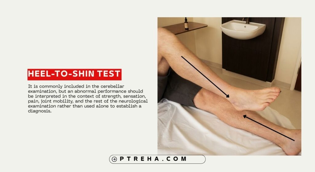
The Heel-to-Shin Test is a bedside neurological assessment used to examine lower-limb coordination and detect features of limb ataxia, particularly dysmetria. It is commonly included in the cerebellar examination, but an abnormal performance should be interpreted in the context of strength, sensation, pain, joint mobility, and the rest of the neurological examination rather than used alone to establish a diagnosis.
What Is the Heel-to-Shin Test?
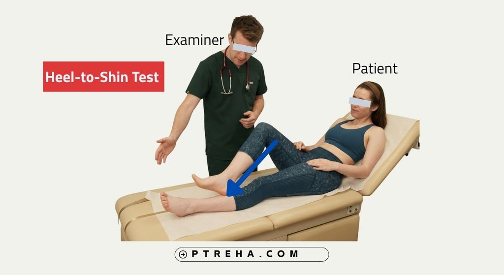
The Heel-to-Shin Test, also called the heel-shin test or heel-shin slide, is a neurological coordination assessment. The patient places the heel of one foot on the opposite knee and slides it down the shin toward the ankle while the examiner observes the accuracy, smoothness, and control of the movement.
The test primarily challenges coordinated movement of the lower limb across multiple joints. Clinically, it is used to look for lower-limb dysmetria and ataxic movement, which may occur with cerebellar dysfunction.
It is important to recognize that the Heel-to-Shin Test is not a standalone diagnostic test for cerebellar disease. Abnormal performance may also be influenced by impaired proprioception, weakness, pain, restricted movement, or other neurological and musculoskeletal factors.
Purpose of the Heel-to-Shin Test
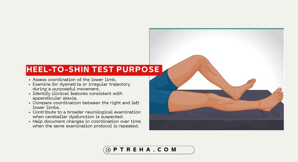
The Heel-to-Shin Test is commonly used to:
- Assess coordination of the lower limb.
- Examine for dysmetria or irregular trajectory during a purposeful movement.
- Identify clinical features consistent with appendicular ataxia.
- Compare coordination between the right and left lower limbs.
- Contribute to a broader neurological examination when cerebellar dysfunction is suspected.
- Help document changes in coordination over time when the same examination protocol is repeated.
It assesses one component of neurological function. It should not be interpreted as a general test of balance, gait, muscle strength, or overall functional mobility.
Heel-to-Shin Test Indications
The assessment may be appropriate when the clinician needs to examine lower-limb coordination, particularly in patients with:
- Suspected or known cerebellar dysfunction.
- Limb ataxia or unexplained incoordination.
- Neurological disorders associated with impaired coordination.
- Changes in gait or motor control accompanied by other cerebellar or neurological findings.
- A need to compare coordination between limbs during a neurological examination.
- A need to monitor coordination as part of a broader rehabilitation assessment.
The indication should come from the patient’s presentation and examination findings rather than from the test being used routinely without a clinical question.
Heel-to-Shin Test Contraindications and Precautions
There are no universally established test-specific contraindications that apply to every patient. Instead, clinicians should consider general safety and the patient’s ability to perform the movement safely.
- Consider pain or an acute musculoskeletal injury that may make the required hip, knee, or ankle movement inappropriate.
- Consider significant weakness that prevents the patient from controlling the limb adequately.
- Take care with severe incoordination or neurological impairment that could make repositioning the limb difficult.
- Ensure that the patient is appropriately supported and positioned on a stable examination surface.
- Modify the procedure when clothing, footwear, or restricted joint movement interferes with the task.
- Stop or modify the examination if the patient develops significant pain, distress, or other concerning symptoms.
These are clinical precautions rather than universal contraindications specific to the Heel-to-Shin Test.
Patient Position
A commonly described protocol uses the patient in a supine position on an examination bed or treatment plinth.
- Position the patient comfortably lying on their back.
- Allow sufficient space for both lower limbs to move freely.
- Remove footwear when it interferes with accurate observation or performance.
- Avoid clothing that restricts movement or obscures the shin.
- Explain the movement before beginning the test.
- Make sure the patient understands which heel should be placed on which knee.
The exact setup can vary between neurological examination protocols.
Examiner Position
- Stand or sit beside the patient’s lower limbs.
- Position yourself so that both legs can be observed throughout the movement.
- Maintain a clear view of the heel, knee, and shin.
- Be prepared to assist the patient if weakness or severe incoordination creates a safety concern.
- Observe both sides using the same instructions and examination conditions.
The clinician should focus on movement quality rather than simply whether the heel reaches the ankle.
How to Perform the Heel-to-Shin Test
A commonly used heel-shin procedure can be performed as follows:
- Explain the task and demonstrate it when necessary.
- Ask the patient to place the heel of the right foot on the left knee.
- Instruct the patient to slide the heel down the front of the left shin toward the ankle.
- Observe whether the heel remains accurately positioned on the shin and whether the movement is smooth and controlled.
- At the distal end of the movement, the patient returns the heel to the opposite knee and repeats the task according to the selected examination protocol.
- Perform the same assessment with the left heel on the right knee.
- Compare the quality of movement between sides.
Some examination protocols use a continuous heel-to-shin movement, whereas formal ataxia assessment instruments may specify a particular number of repetitions or a defined scoring method. Those scoring rules belong to the relevant instrument and should not be treated as universal standards for every Heel-to-Shin Test.
A useful clinical observation is whether the heel remains in contact with the shin and follows a controlled trajectory. Irregular, decomposed, tremulous, or poorly directed movement may indicate impaired lower-limb coordination.
Interpretation
The Heel-to-Shin Test should be interpreted qualitatively unless a validated scoring system is being used.
A well-controlled movement generally shows an accurate trajectory along the opposite shin with appropriate coordination of the limb. In contrast, the clinician may observe difficulty maintaining the heel on the shin, an irregular trajectory, decomposition of movement, or other features of dysmetria.
An abnormal finding can be associated with limb ataxia, including cerebellar dysfunction, but it does not by itself establish the location, cause, or diagnosis of a neurological disorder.
“The quality of the movement is more informative than simply asking whether the heel reached the ankle.”
The clinician should also consider whether the patient has enough strength and joint mobility to perform the task, whether pain is interfering, and whether impaired sensation or proprioception could contribute to the observed performance.
What Does an Abnormal Heel-to-Shin Performance Mean?

An abnormal Heel-to-Shin Test may indicate impaired coordination of the lower limb. When it occurs together with other findings such as dysmetria in the upper limbs, dysarthria, nystagmus, truncal instability, or characteristic gait abnormalities, it may strengthen the clinical suspicion of a cerebellar syndrome.
However, the finding is not specific to one disease or anatomical lesion. Lower-limb incoordination can be influenced by sensory impairment, weakness, pain, restricted range of motion, and other factors that should be assessed separately.
For this reason, the result should be documented descriptively. For example, the clinician may note that the patient demonstrated an irregular heel trajectory, difficulty maintaining contact with the shin, or greater incoordination on one side.
Anatomy Involved in Heel-to-Shin Test
The Heel-to-Shin Test depends on the integrated function of several systems rather than one isolated muscle or anatomical structure.
- Cerebellum: contributes to coordination, timing, movement accuracy, and adaptation of voluntary movement.
- Sensory pathways: proprioceptive information from the lower limb contributes to accurate control of the moving limb.
- Motor pathways: descending motor systems provide the commands required to move and position the limb.
- Hip, knee, and ankle joints: coordinated movement across these joints allows the heel to follow the shin.
- Lower-limb musculature: multiple muscle groups work together to produce and control the movement.
- Visual and other sensory information: can contribute to movement control and should be considered when interpreting performance.
The test therefore reflects integrated neurological and musculoskeletal function rather than isolating a single muscle, nerve, joint, or ligament.
Conditions Associated With the Heel-to-Shin Test
Abnormal performance may be observed in conditions involving impaired coordination, including:
- Cerebellar disorders associated with limb ataxia.
- Hereditary or degenerative ataxias.
- Neurological disorders in which cerebellar signs are present.
- Some disorders causing impaired sensory input or proprioceptive control.
- Other neurological conditions in which lower-limb coordination is impaired.
The Heel-to-Shin Test does not independently identify which condition is present. Diagnosis requires integration of the history, neurological examination, other clinical findings, and, when appropriate, additional investigations.
Clinical Significance
The test can be particularly useful as part of a structured cerebellar and neurological examination.
- It provides information about lower-limb coordination.
- It can reveal side-to-side differences in movement quality.
- It can contribute to recognition of appendicular ataxia.
- It can complement other coordination assessments such as finger-to-nose testing and rapid alternating movements.
- Repeated examination using the same protocol can help clinicians describe changes in performance during rehabilitation or follow-up.
Research examining bedside and instrumented cerebellar tests shows that clinical coordination testing can provide meaningful information about ataxia, but performance depends on the assessment method and the population being examined. Instrumented approaches and formal ataxia scales should therefore not be confused with an informal bedside Heel-to-Shin Test.
Sensitivity and Specificity
A universal sensitivity or specificity value for the Heel-to-Shin Test is not established in a way that supports a single clinically applicable number across populations and protocols.
The available literature includes studies of heel-shin performance within broader cerebellar assessments and studies comparing instrumented coordination tests with ataxia rating scales. These findings do not justify presenting one sensitivity or specificity value as a universal property of the bedside test.
Diagnostic Accuracy
The Heel-to-Shin Test should not be considered a standalone diagnostic test.
Its main clinical value is as one component of the neurological examination. An abnormal result may contribute to the recognition of limb ataxia, but diagnostic interpretation requires integration with the patient’s history, other neurological signs, and appropriate investigations.
Common Mistakes in Heel-to-Shin Test
- Treating the test as a standalone test for cerebellar disease.
- Using a universal pass/fail rule that is not part of the selected protocol.
- Focusing only on whether the heel reaches the ankle.
- Failing to compare both lower limbs.
- Ignoring pain, weakness, restricted mobility, or sensory impairment.
- Giving inconsistent instructions between sides.
- Allowing clothing or footwear to interfere with the movement.
- Testing a severely impaired patient without considering appropriate support.
- Mistaking an abnormal movement for proof of a specific neurological diagnosis.
- Changing the protocol between assessments and then interpreting the results as directly comparable.
Tips for Performing the Heel-to-Shin Test
- Explain and demonstrate the movement before testing.
- Use consistent instructions on both sides.
- Observe the entire movement rather than only its endpoint.
- Look at accuracy, trajectory, rhythm, smoothness, and the ability to maintain contact with the shin.
- Compare sides without assuming that asymmetry automatically identifies the cause.
- Consider strength, sensation, pain, joint mobility, and cognition before interpreting an abnormal performance.
- Document the actual observed finding rather than simply writing “positive” or “negative.”
- Use a formal ataxia rating scale when a standardized score is clinically required rather than inventing a bedside score.
- When monitoring change over time, reproduce the same protocol and testing conditions as closely as practical.
“An abnormal Heel-to-Shin Test is a clinical finding, not a diagnosis.”
Summary
- The Heel-to-Shin Test is a neurological clinical assessment of lower-limb coordination and is commonly used when examining for limb ataxia.
- The patient typically places one heel on the opposite knee and slides it along the shin toward the ankle while the clinician observes movement quality.
- Difficulty maintaining the heel on the shin, an irregular trajectory, or poorly coordinated movement may indicate impaired lower-limb coordination.
- Abnormal performance can be associated with cerebellar dysfunction but may also be influenced by sensory impairment, weakness, pain, or musculoskeletal limitations.
- The test should be interpreted alongside the patient’s history and the broader neurological examination rather than used as a standalone diagnostic tool.
The Heel-to-Shin Test remains a simple and useful component of bedside neurological examination when performed consistently and interpreted in context. Its greatest value comes from careful observation of movement quality and integration of the finding with the rest of the clinical assessment.
Resources and External Links
Peer-Reviewed and Academic Resources
Power L, Pathirana P, Horne M, et al. Instrumented Objective Clinical Examination of Cerebellar Ataxia: the Upper and Lower Limb—a Review. Cerebellum. This review discusses bedside and instrumented assessments of cerebellar ataxia, including the heel-shin examination.
PubMed – Instrumented Objective Clinical Examination of Cerebellar Ataxia
Perez-Lloret S, van de Warrenburg BP, Rossi M, et al. Assessment of Ataxia Rating Scales and Cerebellar Functional Tests: Critique and Recommendations. Movement Disorders. This review discusses clinical assessment tools used in cerebellar disorders and their appropriate interpretation.
PubMed – Assessment of Ataxia Rating Scales and Cerebellar Functional Tests
Kuo SH. Ataxia. Continuum: Lifelong Learning in Neurology. A clinical review of cerebellar ataxia and associated neurological findings.
PMC – Ataxia
Evaluation of Cerebellar Ataxic Patients. This clinical review describes examination of appendicular ataxia, including the heel-shin slide.
PMC – Evaluation of Cerebellar Ataxic Patients
Clinical Methods: The Cerebellum. NCBI Bookshelf provides a traditional neurological examination description of heel-shin testing and cerebellar assessment.
NCBI Bookshelf – The Cerebellum
Cerebellar Dysfunction. StatPearls, NCBI Bookshelf, provides an overview of cerebellar examination findings and clinical interpretation of the heel-shin test.
NCBI Bookshelf – Cerebellar Dysfunction