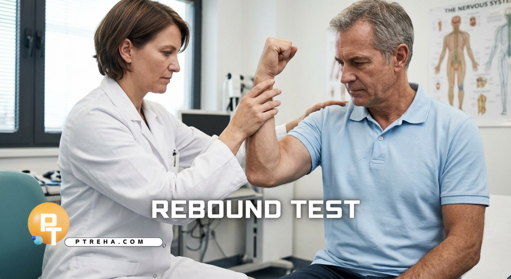
The Rebound Test, also called the Stewart–Holmes maneuver or Stewart holmes test, is a neurological bedside assessment used during examination of motor control and cerebellar function.
It challenges the patient’s ability to control a limb when resistance is suddenly removed, making it particularly useful for observing impaired braking or “checking” of movement.
What Is the Rebound Test?
The Rebound Test is a neurological clinical assessment, not a functional performance test or standardized outcome measure.
It is traditionally included within the examination of cerebellar motor function and assesses how effectively the nervous system coordinates agonist and antagonist muscle activity during a sudden change in resistance.
In the classic maneuver, the patient produces a voluntary movement against the examiner’s resistance.
When that resistance is released unexpectedly, a healthy response includes a controlled movement followed by rapid braking by the opposing musculature.
In cerebellar dysfunction, the braking response may be impaired, allowing excessive movement or overshoot.
An important terminology point is that the rebound phenomenon itself is not inherently abnormal.
Holmes described a rebound response in unaffected limbs, an exaggerated response in spastic limbs, and an absent rebound response in limbs affected by cerebellar disease.
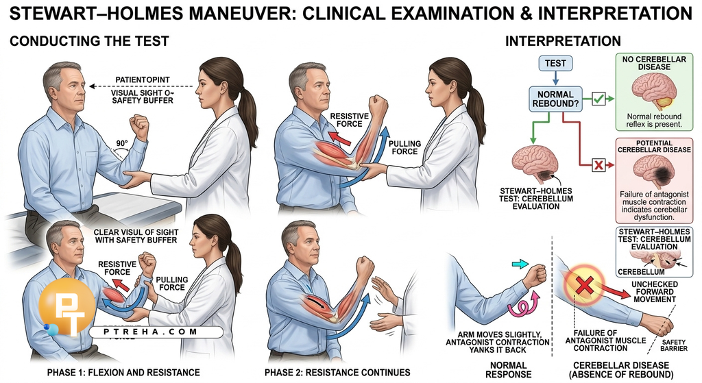
Different clinical descriptions use slightly different upper-limb techniques.
The procedure below describes a commonly taught seated version in which the patient flexes the elbow against resistance.
Purpose of the Stewart holmes test
The Rebound Test may be used to:
- Examine the ability to rapidly check or brake a voluntary movement.
- Identify an abnormal rebound response during a neurological examination.
- Provide clinical information relevant to cerebellar motor dysfunction.
- Complement other assessments of coordination, tone, dysmetria, and motor control.
The maneuver should be considered one component of a broader neurological examination rather than an isolated test of cerebellar function.
Rebound Test Indications
The test may be considered when the clinical examination raises questions about:
- Possible cerebellar dysfunction or cerebellar ataxia.
- Abnormal limb coordination or dysmetria.
- Impaired control of rapid changes in movement.
- Abnormal muscle tone, particularly when distinguishing cerebellar findings from exaggerated responses associated with spasticity.
It can be particularly informative when interpreted alongside finger-to-nose testing, rapid alternating movements, gait assessment, eye-movement examination, and assessment of muscle tone.
Rebound Test Contraindications and Precautions
A universally accepted list of test-specific contraindications is not established for this bedside neurological maneuver.
Clinical judgment should determine whether the movement is appropriate for the individual patient.
Relevant precautions include:
- Avoid applying excessive resistance or an unnecessarily forceful release.
- Protect the patient’s face because an impaired braking response can allow the forearm to move rapidly toward the head.
- Perform the maneuver with the patient seated when this provides greater safety and control.
- Consider pain, acute joint injury, marked weakness, or significant musculoskeletal instability before asking the patient to resist force.
- Stop the test if the patient develops pain, distress, loss of balance, or an unsafe movement.
The examiner should position the other hand or body so that an unexpectedly large movement cannot cause facial or other injury.
Standard neurological examination references specifically emphasize preventing the tested arm from striking the patient’s face.
Patient Position
A commonly described position is:
- Patient seated comfortably with the trunk supported and the tested upper limb accessible.
- Shoulder positioned so the examiner can control the limb safely.
- Elbow flexed while the patient prepares to generate force against the examiner.
- Patient instructed clearly to maintain the requested position or movement.
The exact joint angle and hand position can vary between teaching protocols.
The important feature is that the patient can voluntarily generate movement against controlled resistance while the examiner can safely release that resistance.
Examiner Position
- Stand or sit facing the patient on the side being examined.
- Maintain close control of the patient’s tested limb.
- Use one hand to provide resistance at the distal limb according to the selected protocol.
- Keep the opposite hand or another protective position ready to prevent excessive movement toward the patient’s face.
- Observe the entire movement rather than focusing only on the final position.
Good examiner positioning is essential because the test intentionally creates a sudden change in resistance.
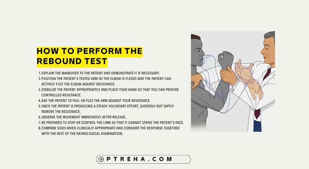
How to Perform the Rebound Test
Using the classic upper-limb version:
- Explain the maneuver to the patient and demonstrate it if necessary.
- Position the patient’s tested arm so the elbow is flexed and the patient can actively flex the elbow against resistance.
- Stabilize the patient appropriately and place your hand so that you can provide controlled resistance.
- Ask the patient to pull or flex the arm against your resistance.
- Once the patient is producing a steady voluntary effort, suddenly but safely remove the resistance.
- Observe the movement immediately after release.
- Be prepared to stop or control the limb so that it cannot strike the patient’s face.
- Compare sides when clinically appropriate and consider the response together with the rest of the neurological examination.
The release should be sufficiently sudden to challenge the braking response, but it should not be unnecessarily forceful.
The objective is to observe motor control, not to generate a large mechanical movement.
“The important observation is how effectively the limb is checked after resistance disappears.”
Test Findings and Interpretation
Because terminology surrounding the Rebound Test can be confusing, describing the observed response is preferable to simply labeling a maneuver “positive” or “negative.”
A normal response includes some movement following release, followed by effective action of the antagonist muscles that checks the movement.
The limb should not continue moving uncontrollably through a large excursion.
An abnormal response may involve excessive rebound or failure to adequately check the movement.
In cerebellar dysfunction, the patient may be unable to decelerate the limb effectively, producing marked overshoot.
This phenomenon is commonly discussed as a manifestation of impaired coordination or dysynergia.
An important distinction is that the direction and degree of abnormality are not interpreted in isolation.
An exaggerated response can occur with spasticity, whereas the classical description associates an absent rebound response with cerebellar disease.
Therefore, the clinician should document what was actually observed—for example, excessive rebound, marked overshoot, or poor checking of movement—rather than relying on an unexplained positive/negative label.
What Does an Abnormal Rebound Response Mean?
An abnormal Rebound Test finding can indicate impaired control of a moving limb, particularly difficulty coordinating agonist and antagonist muscle activity after the external resistance changes.
In the appropriate clinical context, excessive rebound or failure of checking may support the presence of cerebellar motor dysfunction.
However, the finding does not by itself establish a cerebellar lesion or a specific neurological diagnosis.
It should be interpreted alongside other examination findings and the patient’s history.
Spasticity can produce an exaggerated rebound response through a different neurological mechanism, which is one reason the test should not be interpreted as a simple cerebellar “yes or no” test.
“An abnormal rebound response is a clinical sign, not a diagnosis.”
Anatomy Involved in Rebound Test
The maneuver depends on interaction among several parts of the motor system:
- Cerebellum: contributes to coordination, timing, and appropriate scaling of muscular forces during movement.
- Cerebellar motor pathways: participate in the coordination required to modify movement rapidly.
- Motor cortex and descending pathways: generate and control the voluntary effort against resistance.
- Spinal and peripheral motor systems: execute the muscular response required to produce and check movement.
- Agonist and antagonist muscles: alternate their contributions to produce and then brake the movement.
The test therefore reflects integrated neurological motor control rather than the isolated function of one muscle, tendon, peripheral nerve, or joint.
The cerebellum is particularly relevant because impaired checking and rebound are recognized features of cerebellar motor syndromes.
Conditions Detected / Associated With the Test
The Rebound Test can contribute to assessment of:
- Cerebellar dysfunction or cerebellar ataxia, particularly when accompanied by other cerebellar signs.
- Neurological disorders involving cerebellar motor control.
- Spastic motor syndromes, where an exaggerated rebound response may occur.
The test does not independently identify the underlying cause of cerebellar dysfunction.
Conditions such as stroke, demyelinating disease, degenerative disorders, tumors, intoxication, or other neurological disorders require appropriate history, neurological examination, and further investigation when indicated.
Clinical Significance
The Rebound Test can be useful because it examines a specific aspect of movement control that may not be obvious during ordinary voluntary movement.
Clinically, it can:
- Add information to a cerebellar examination.
- Help reveal impaired deceleration or checking of movement.
- Contribute to neurological localization when interpreted with other findings.
- Help characterize abnormal motor responses when tone abnormalities are also present.
- Provide a rapid bedside observation without requiring specialized equipment.
Its main value is therefore contextual.
A markedly abnormal response becomes more meaningful when it occurs together with other findings such as dysmetria, dysdiadochokinesia, gait ataxia, hypotonia, or abnormal eye movements.
Sensitivity and Specificity
Reliable universal sensitivity and specificity values for the bedside Rebound Test are not established in a way that supports a single general cutoff or diagnostic accuracy claim.
The maneuver is primarily used as part of the neurological examination, and its interpretation depends on the clinical context and the method used.
It should not be presented as a standalone diagnostic test with a universal sensitivity or specificity value.
Diagnostic Accuracy
A universally applicable diagnostic-accuracy estimate for the Rebound Test is not established.
The test is better understood as a clinical sign within neurological examination than as an independent diagnostic procedure.
Modern assessment of cerebellar dysfunction generally relies on a combination of examination findings rather than one maneuver alone.
Common Mistakes in Rebound Test
- Applying excessive resistance before release.
- Failing to protect the patient’s face from the moving limb.
- Releasing the resistance without first establishing a clear voluntary effort.
- Interpreting every rebound response as abnormal.
- Calling an exaggerated response evidence of cerebellar disease without considering spasticity.
- Using “positive” and “negative” labels without describing the actual observed movement.
- Treating the maneuver as a standalone diagnostic test.
- Ignoring pain, weakness, joint pathology, or poor patient understanding that may alter performance.
- Failing to compare the finding with other neurological examination results.
Tips for Performing the Rebound Test
- Explain exactly what the patient should do before applying resistance.
- Use enough resistance to create a controlled challenge, not enough to overpower the patient.
- Keep the release sudden but mechanically safe.
- Protect the patient’s face throughout the maneuver.
- Observe the first movement immediately after release.
- Compare sides when appropriate.
- Describe the response precisely rather than relying only on “positive” or “negative.”
- Interpret the finding in relation to tone, strength, coordination, gait, and other cerebellar signs.
- Repeat the maneuver only when necessary and when doing so remains safe.
Summary
The Rebound Test is a neurological clinical assessment of movement checking and motor coordination, traditionally associated with the Stewart–Holmes maneuver.
It is performed by asking the patient to generate movement against resistance and then suddenly removing that resistance while the examiner observes the patient’s ability to control the resulting movement.
Excessive rebound or poor checking can be associated with cerebellar dysfunction, while exaggerated responses may also occur with spasticity.
The response should be interpreted as one finding within a complete neurological examination rather than as a standalone diagnostic test.
There is no universally accepted sensitivity, specificity, cutoff, or diagnostic-accuracy value that should be applied to the maneuver across all patients and protocols.
When performed correctly, the Rebound Test provides a simple bedside observation of how effectively the nervous system controls and brakes movement after a sudden change in resistance.
Resources and External Links
1.
Angel RW.
The Rebound Phenomenon of Gordon Holmes.
Archives of Neurology.
A classic discussion clarifying that rebound is normally present, may be exaggerated with spasticity, and may be absent with cerebellar disease.
PubMed – The Rebound Phenomenon of Gordon Holmes
2.
Manto M, et al.
Consensus Paper: Revisiting the Symptoms and Signs of Cerebellar Syndrome.
Cerebellum.
A consensus review describing the Stewart–Holmes maneuver among clinical maneuvers used to evaluate cerebellar motor syndrome.
PMC – Consensus Paper: Revisiting the Symptoms and Signs of Cerebellar Syndrome
3.
Clinical Methods: The Cerebellum – NCBI Bookshelf.
A clinical neurological examination reference describing the upper-limb rebound maneuver and the importance of protecting the patient from excessive arm movement.
NCBI Bookshelf – The Cerebellum
4.
Cerebellar Dysfunction – StatPearls, NCBI Bookshelf.
A clinical overview that includes the rebound phenomenon among examination findings associated with cerebellar dysfunction.
NCBI Bookshelf – Cerebellar Dysfunction
5.
Holmes GM.
The Symptoms of Acute Cerebellar Injuries Due to Gunshot Injuries.
Brain.
The historical neurological work associated with Holmes’ description of cerebellar signs, including the rebound phenomenon.