
Homan’s Sign is a historical pain-provocation finding associated with calf discomfort during passive ankle dorsiflexion. It cannot reliably confirm or exclude deep vein thrombosis (DVT), and suspected DVT requires a clinical diagnostic pathway rather than repeated attempts to elicit this sign.
What is Homan’s Sign?
Homan’s Sign, also written Homans’ sign, is traditionally described as calf discomfort when the foot is passively moved upward at the ankle. It is best classified as a historical clinical sign, not a validated stand-alone DVT screening test.
A clinical study comparing symptoms and signs with objective venous testing found that classic findings, including Homan’s Sign, occurred with similar frequency in limbs with and without DVT in its referred sample. This does not support using the sign to decide whether a clot is present.
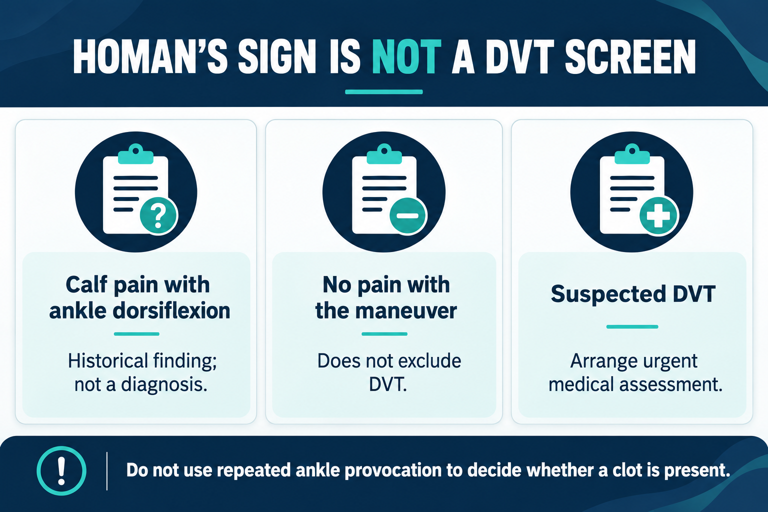
Historical purpose and present clinical role
- The maneuver was used to look for calf pain thought to accompany venous thrombosis.
- A painful response is nonspecific.
- A painless response does not exclude a clot.
- Its useful teaching role is to explain the limitations of isolated bedside signs.
There is no reason to wait for Homan’s Sign before escalating a suspected DVT. It should not be used to clear a patient for massage, exercise, or discharge.
Safety, precautions, and when to defer provocation
When DVT is suspected, do not use forceful or repeated ankle provocation to obtain reassurance. Arrange the appropriate medical assessment and investigations instead.
- Do not delay referral while trying to produce a positive finding.
- Avoid painful passive movement in an acutely injured, fractured, or postoperative limb when movement is restricted.
- Respect consent and existing joint or tissue precautions.
- Do not encourage patients to perform the maneuver as a home clot check.
The response does not safely guide DVT decisions. Suspected thrombosis needs a diagnostic pathway even when passive ankle movement is comfortable.
Patient position and the historical maneuver
A commonly described variant uses a patient lying supine with the knee extended and the examiner positioned at the foot. The description below explains the historical finding; it is not a recommendation to use it in a suspected-DVT examination.
- The lower limb is supported with the knee extended.
- The examiner passively dorsiflexes the ankle, moving the foot toward the shin.
- Calf discomfort is described as the historical positive response.
- The response is documented without using it to confirm or exclude thrombosis.
Descriptions vary, so findings from different techniques should not be treated as equivalent. There is no validated force or ankle-angle target that turns this sign into a reliable clot test.
Positive and negative findings: what they mean
A “positive” Homan’s Sign describes pain provoked during the maneuver, not a positive laboratory or imaging diagnosis of DVT. A “negative” sign means pain was not elicited under the conditions used.
- Positive: pain occurred; the cause still requires assessment.
- Negative: pain was absent; DVT remains possible.
- Untested: the maneuver was not performed; this is not a missing requirement for a DVT work-up.
- Limited by pain or stiffness: record the limitation without assigning diagnostic certainty.
Do not use either result as a rule-in or rule-out decision. History, clinical probability, and appropriate investigations carry the decision-making role.
Relevant anatomy and alternative causes of calf pain
- DVT involves the deep venous system, commonly in the lower limb.
- Ankle dorsiflexion changes tension in calf and posterior ankle tissues.
- The maneuver does not isolate a vein, identify a thrombus, or locate its extent.
- Musculoskeletal conditions and popliteal cyst-related symptoms can also resemble a painful calf presentation.
Local tissue pain and venous disease cannot be reliably separated by this one movement. A plausible alternative explanation does not automatically exclude DVT when the clinical picture remains concerning.
What should replace Homan’s Sign in suspected DVT?
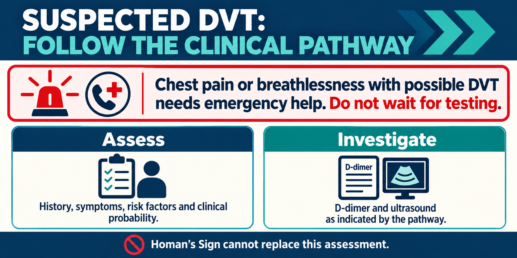
Use a validated local pathway. The NICE recommendations for suspected DVT in adults combine history and examination with clinical probability assessment, D-dimer testing, and proximal leg vein ultrasound as appropriate.
- Assess symptom onset, swelling, pain, relevant medical history, and risk factors.
- Use the clinical probability tool required by the pathway in the population for which it applies.
- Arrange D-dimer and ultrasound in the indicated sequence.
- Follow the pathway when further or repeat testing is required.
- Leave anticoagulation decisions to the responsible prescribing team.
A probability score is not itself proof of thrombosis. Likewise, an isolated D-dimer result should not be interpreted outside the relevant diagnostic pathway.
Urgent symptoms and escalation
Possible DVT symptoms include new pain and swelling in a leg, sometimes with skin colour change. Seek urgent medical assessment rather than testing the ankle at home.
- New unexplained one-sided leg pain or swelling warrants prompt assessment.
- Chest pain or breathlessness alongside possible DVT symptoms requires emergency help.
- Tell the receiving team about recent surgery, reduced mobility, previous clots, and other relevant history.
The NHS guidance on DVT symptoms and emergency signs explains why chest symptoms must not be ignored. A negative Homan’s Sign should never downgrade that urgency.
Diagnostic accuracy and practical mistakes
The evidence does not justify a universal sensitivity or specificity estimate for routine use across populations and techniques. Neither the historical association nor a remembered numerical estimate makes Homan’s Sign an adequate screening test.
- Do not mistake a historical “positive sign” for confirmed disease.
- Do not record “DVT excluded” because dorsiflexion was painless.
- Do not substitute repeated calf or ankle provocation for referral.
- Do not use the sign to choose treatment for an undiagnosed painful calf.
For practical documentation, record the symptoms and actions taken. For example, describing unilateral swelling and arranging urgent assessment is more informative than documenting a reassuring ankle response.
Summary
- Homan’s Sign is a historical pain-provocation finding, not a dependable DVT screen.
- Calf discomfort during passive ankle dorsiflexion is nonspecific.
- A painless maneuver does not exclude DVT.
- Suspected DVT needs a validated diagnostic pathway and appropriate testing.
- Possible DVT with chest pain or breathlessness requires emergency escalation.
The useful clinical lesson is to recognize the sign’s limitations and act on the whole presentation. Timely assessment matters more than reproducing or dismissing calf pain with ankle movement.