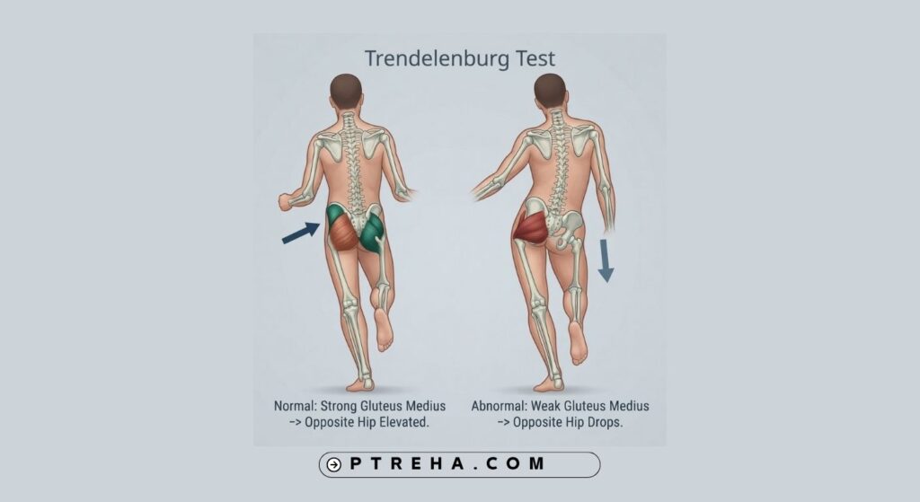
The Trendelenburg Test is a clinical assessment used to observe how well the hip abductor mechanism stabilizes the pelvis during single-leg stance. A drop of the pelvis on the unsupported side can indicate impaired ability of the weight-bearing hip to maintain pelvic stability, but the finding should be interpreted alongside the patient’s symptoms, strength, range of motion, gait, and broader clinical examination.
What Is the Trendelenburg Test?
The Trendelenburg Test is a clinical physical examination assessment, rather than a standalone diagnostic test. It is commonly used to assess pelvic control during single-leg stance and to identify a possible deficit in the hip abductor mechanism.
During the assessment, the patient stands on one leg while the examiner observes the position of the pelvis. Normally, the weight-bearing hip abductors contribute to maintaining the pelvis in a relatively stable position. If the pelvis drops toward the side of the lifted leg, this is traditionally described as a positive Trendelenburg sign.
The finding is commonly associated with reduced function of the hip abductors, particularly the gluteus medius and gluteus minimus. However, pelvic control during single-leg stance is influenced by more than one muscle or structure, so the test should not be interpreted as an isolated test of a single muscle.
“A pelvic drop is a clinical finding, not a diagnosis.”
The Trendelenburg assessment should therefore be considered one component of a comprehensive musculoskeletal or neurological examination.
Purpose of the Trendelenburg Test
The assessment may be used to:
- Observe pelvic stability during single-leg stance.
- Identify a possible deficit in the hip abductor mechanism.
- Examine asymmetry between the two lower limbs.
- Support the clinical assessment of patients with hip-related symptoms.
- Observe a feature that may also appear during gait.
- Help guide further examination of hip strength, motor control, pain, and functional movement.
- Monitor changes in pelvic control during rehabilitation when the test is repeated under a consistent protocol.
The Trendelenburg finding may occur in several different clinical situations, including conditions affecting the hip abductor mechanism, painful hip disorders, neurological problems, and some structural disorders of the hip. It therefore does not identify one specific underlying pathology by itself.
Trendelenburg Test Indications
The assessment may be considered when a clinician is evaluating:
- Suspected weakness or impaired function of the hip abductors.
- Abnormal pelvic control during walking.
- A lateral trunk lean or suspected Trendelenburg gait.
- Hip pain accompanied by apparent difficulty maintaining pelvic stability.
- Functional asymmetry between the lower limbs.
- Hip disorders in which abductor function may be clinically relevant.
- Possible neurological involvement affecting the hip abductor mechanism.
- Changes in hip function following relevant injury or surgery.
The test can be particularly useful when combined with gait observation and manual or instrumented assessment of hip muscle performance.
Trendelenburg Test Contraindications and Precautions
There is no single universally established list of test-specific contraindications. Clinical judgment is required because the assessment requires the patient to stand on one lower limb.
Important safety precautions include:
- Do not perform the test without appropriate support when the patient has poor balance or a significant risk of falling.
- Avoid forcing a patient into single-leg stance when doing so produces significant pain or instability.
- Consider whether the patient can understand and follow the instructions.
- Consider whether the patient has sufficient lower-limb control to perform the task safely.
- Be cautious when substantial hip, pelvic, or lower-limb pain prevents meaningful weight-bearing.
- Use an appropriate guarding position and allow the patient to regain two-leg support immediately if balance is lost.
Pain and poor balance can interfere with the test and may produce an apparent pelvic deviation that does not simply reflect hip abductor weakness.
Patient Position
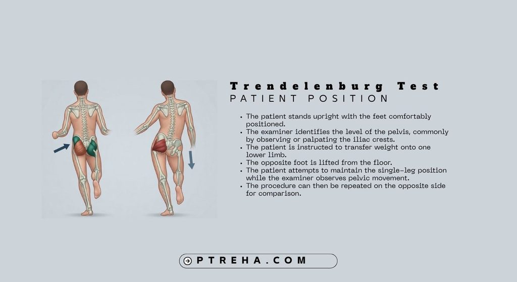
For a commonly used standing version of the assessment:
- The patient stands upright with the feet comfortably positioned.
- The examiner identifies the level of the pelvis, commonly by observing or palpating the iliac crests.
- The patient is instructed to transfer weight onto one lower limb.
- The opposite foot is lifted from the floor.
- The patient attempts to maintain the single-leg position while the examiner observes pelvic movement.
- The procedure can then be repeated on the opposite side for comparison.
The patient should be positioned so that clothing and environmental factors do not obscure observation of pelvic movement.
Examiner Position
The examiner commonly:
- Stands behind the patient.
- Positions themselves close enough to observe pelvic movement clearly.
- Maintains an appropriate guarding position to reduce fall risk.
- Observes the pelvis from behind rather than relying only on the patient’s subjective sensation.
- May lightly palpate the iliac crests to help identify changes in pelvic level.
- Compares the response between the right and left sides.
A stable and consistent examiner position is important because small changes in viewing angle can influence visual interpretation.
How to Perform the Trendelenburg Test
A practical clinical procedure is:
- Explain the procedure to the patient and ensure that the patient understands the task.
- Position the patient in standing.
- Stand behind the patient in a safe guarding position.
- Observe the pelvis before single-leg stance.
- Ask the patient to transfer their weight onto one leg.
- Ask the patient to lift the opposite foot from the floor.
- Observe whether the pelvis remains relatively level or drops toward the unsupported side.
- Observe any compensatory movement of the trunk.
- Return the patient to double-leg stance.
- Repeat on the opposite side.
- Compare the two sides and relate the finding to the remainder of the examination.
The exact duration and instructions can vary between clinical protocols. Therefore, a specific duration should not be treated as a universal diagnostic threshold.
Some clinical descriptions use a sustained single-leg stance, while other approaches emphasize observation of pelvic control during gait.
Interpretation of the Trendelenburg Test
The traditional interpretation focuses on the relationship between the stance limb and the unsupported side.
If the pelvis drops toward the side of the lifted leg while the patient is standing on the opposite limb, the finding is traditionally called a positive Trendelenburg sign. This suggests that the weight-bearing hip abductor mechanism may not be adequately controlling the pelvis.
For example, if the patient stands on the right leg and the left side of the pelvis drops, the finding suggests impaired pelvic stabilization associated with the right stance limb.
However, this observation does not establish that the gluteus medius is the sole cause. Hip abductor performance can be affected by muscle weakness, pain, altered mechanics, neurological impairment, structural changes, or other factors.
The examiner should therefore ask:
- Is the finding reproducible?
- Is it present on one side or both?
- Does it occur because of pain?
- Is there an obvious trunk compensation?
- Is hip abduction strength also impaired?
- Is the patient’s gait affected?
- Are there neurological findings?
- Are there structural or range-of-motion abnormalities that could influence the result?
“Interpret the pelvic movement in the context of the whole examination.”
What Does a Positive Trendelenburg Test Mean?
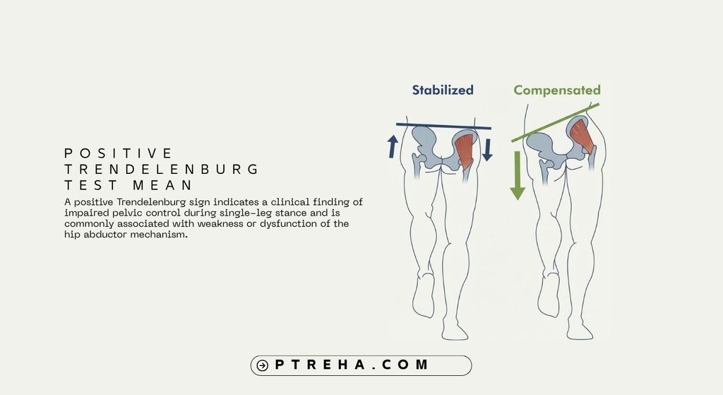
A positive Trendelenburg sign indicates a clinical finding of impaired pelvic control during single-leg stance and is commonly associated with weakness or dysfunction of the hip abductor mechanism.
It does not, by itself, establish a specific diagnosis.
Potential contributors include:
- Reduced hip abductor muscle performance.
- Pain-related inhibition.
- Disorders affecting the hip or proximal femur.
- Injury or dysfunction of the superior gluteal nerve.
- Neuromuscular conditions.
- Structural changes affecting the mechanical function of the hip abductors.
- Postoperative changes affecting the abductor mechanism.
The clinical context determines which of these possibilities is relevant. Further assessment is required before attributing the finding to a particular pathology.
Negative Trendelenburg Test
When the pelvis remains adequately controlled during single-leg stance, the clinician may describe the test as negative according to the traditional terminology.
However, an apparently normal response does not exclude hip pathology, weakness, pain, or other movement impairments. A patient may demonstrate adequate pelvic control during a relatively simple single-leg task while showing abnormalities during walking, running, stair negotiation, or other higher-demand activities.
The result should therefore not be treated as a definitive rule-out test.
Anatomy Involved in Trendelenburg Test
The main anatomical and physiological components relevant to the assessment include:
- Gluteus medius: an important hip abductor and contributor to pelvic stabilization during weight-bearing.
- Gluteus minimus: contributes to hip abduction and hip stabilization.
- Tensor fasciae latae: also contributes to hip abduction and can influence frontal-plane hip control.
- Superior gluteal nerve: supplies the gluteus medius, gluteus minimus, and tensor fasciae latae.
- Pelvis: its movement provides the principal observable outcome during the standing assessment.
- Hip joint and proximal femur: their structure and mechanical relationship influence the effectiveness of the abductor mechanism.
The gluteus medius and minimus are important contributors, but the Trendelenburg Test should not be regarded as an isolated test of either muscle.
Conditions Associated With the Test
A Trendelenburg sign can be associated with a variety of conditions rather than one specific diagnosis.
Examples include:
- Hip abductor weakness or dysfunction.
- Disorders affecting the hip abductor mechanism.
- Developmental or structural hip disorders.
- Certain neurological disorders affecting the superior gluteal nerve or its pathways.
- Painful hip conditions that interfere with effective abductor function.
- Some postoperative hip conditions.
- Conditions involving the greater trochanter or proximal femur.
A Trendelenburg finding has also been described in patients with greater trochanteric pain syndrome, although the finding should be interpreted together with the patient’s pain location, symptom behavior, strength findings, and other examination findings.
Clinical Significance
The Trendelenburg Test can provide useful information about frontal-plane pelvic control, but its greatest value comes from combining it with other clinical findings.
Clinicians may use the result to:
- Decide whether more detailed hip abductor assessment is warranted.
- Compare functional performance between limbs.
- Identify movement compensations.
- Support gait analysis.
- Help formulate rehabilitation goals.
- Monitor changes in pelvic control over time.
- Determine whether additional neurological, musculoskeletal, or structural assessment is appropriate.
A trunk lean toward the stance side may occur as a compensation because shifting the trunk can alter the mechanical demand placed on the hip abductors. This can make the pelvic movement appear less pronounced and should be documented rather than ignored.
Sensitivity and Specificity
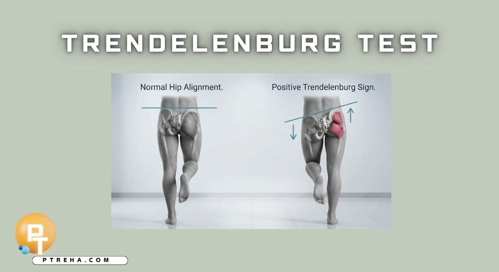
A single universally applicable sensitivity or specificity value should not be assigned to the Trendelenburg Test.
Reported performance can depend on the population studied, the exact test protocol, the definition of an abnormal finding, and the reference standard. In addition, pelvic drop does not necessarily correspond directly to measured hip abductor strength in every population.
For this reason, the Trendelenburg Test should be considered a clinical examination finding rather than a standalone diagnostic accuracy test.
Diagnostic Accuracy
The Trendelenburg Test should not be used alone to establish a diagnosis.
A pelvic drop may indicate impaired hip abductor function, but similar movement findings can arise from different mechanisms. Conversely, a patient with clinically relevant hip pathology may not demonstrate an obvious Trendelenburg sign during a particular examination.
A complete assessment may therefore include:
- Subjective history.
- Pain assessment.
- Observation of standing posture.
- Hip range of motion.
- Hip muscle performance.
- Neurological examination when indicated.
- Gait analysis.
- Functional movement assessment.
- Additional orthopedic or medical investigations when clinically justified.
The available clinical literature emphasizes that the Trendelenburg sign contributes to assessment but does not independently establish the underlying hip disorder.
Common Mistakes in Trendelenburg Test
Common clinical errors include:
- Calling every pelvic asymmetry a positive Trendelenburg sign.
- Assuming that a positive finding automatically means isolated gluteus medius weakness.
- Ignoring pain during single-leg stance.
- Failing to assess balance before performing the test.
- Allowing excessive trunk movement without documenting the compensation.
- Observing only one limb.
- Relying on the test without performing a broader hip and gait examination.
- Treating the result as a definitive diagnosis.
- Using inconsistent instructions between repeated assessments.
- Ignoring structural or neurological factors that may influence pelvic control.
One particularly important mistake is confusing the Trendelenburg sign with Trendelenburg gait. The sign is observed during a clinical single-leg stance assessment, whereas Trendelenburg gait refers to a gait pattern characterized by impaired pelvic control associated with the stance limb.
Tips for Performing the Trendelenburg Test
For more useful clinical observation:
- Perform the assessment in a safe environment.
- Expose or otherwise visualize the pelvic landmarks adequately.
- Use the same instructions when comparing both sides.
- Observe the pelvis from behind.
- Look for trunk compensation as well as pelvic movement.
- Ask about pain rather than assuming that movement loss represents weakness.
- Compare the right and left sides.
- Combine the finding with resisted hip abduction or other appropriate strength assessment.
- Consider gait observation when the clinical question involves walking.
- Document the actual observed movement rather than simply recording “positive” or “negative.”
- Repeat the assessment consistently when using it to monitor rehabilitation progress.
Limitations of the Trendelenburg Test
The Trendelenburg Test provides information about pelvic control under a particular task. It does not directly quantify overall hip strength, balance, gait quality, or functional capacity.
Pelvic movement can also be influenced by pain, balance, compensatory strategies, structural alignment, and the patient’s ability to perform the task. Published clinical discussions have also noted that pelvic drop does not always correspond directly with hip abductor strength.
Therefore, a clinician should avoid making a diagnosis or treatment decision based solely on the Trendelenburg finding.
Summary
- The Trendelenburg Test is a clinical assessment of pelvic control during single-leg stance.
- A drop of the pelvis toward the unsupported side is traditionally described as a positive Trendelenburg sign.
- The finding is commonly associated with impaired function of the weight-bearing hip abductor mechanism, particularly the gluteal abductors.
- A positive finding does not identify a specific diagnosis or prove isolated weakness of one muscle.
- Pain, balance, structural factors, neurological impairment, and compensatory movement can influence the result.
- The assessment is most useful when combined with history, strength testing, range-of-motion assessment, gait analysis, and other relevant clinical findings.
The Trendelenburg Test remains a simple and useful component of the physical examination, particularly when the clinician is evaluating hip function and pelvic stability. Its value comes from careful observation and appropriate clinical interpretation rather than treating the finding as a standalone diagnostic answer.