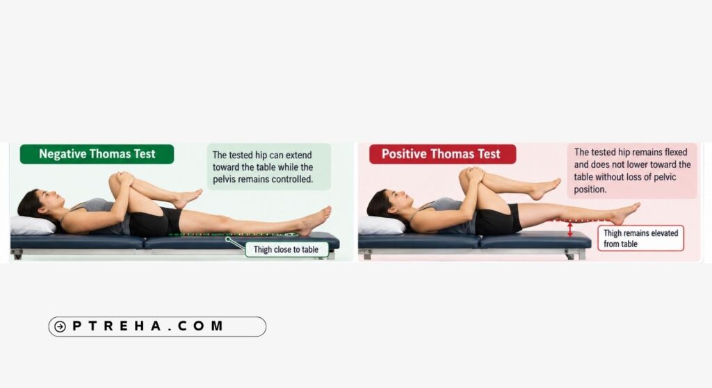
The Thomas Test is a commonly used clinical assessment for identifying limited hip extension associated with hip flexion tightness or contracture.
Its value lies in observing hip and pelvic behavior under controlled positioning, not in diagnosing a specific muscle disorder or establishing the cause of a patient’s symptoms by itself.
What Is the Thomas Test?
The Thomas Test is a physical examination maneuver used to assess whether a hip can extend toward a neutral position while the opposite hip is flexed to control the pelvis and lumbar spine.
It is most commonly used when a clinician suspects a hip flexion contracture or restriction in hip extension.
The classic Thomas Test should be distinguished from the Modified Thomas Test.
In the modified version, the patient is usually positioned near the edge of the examination table so that the tested thigh and lower leg can move more freely.
Both variations are frequently discussed in relation to hip-flexor flexibility, but positioning and interpretation differ between protocols.
From a physical therapy examination standpoint, the Thomas Test should normally be one component of a broader assessment that may include:
- Hip range of motion
- Pelvic position
- Lumbar spine movement
- Gait observation
- Functional movements
- Muscle performance
- Symptom behavior
- Relevant orthopedic examination findings
A hip examination review published in the medical literature describes the Thomas maneuver as a method of assessing hip flexion tightness or contracture while emphasizing the importance of controlling pelvic position during the examination.
“The quality of pelvic control during the Thomas Test is often as important as the apparent position of the thigh.”
Purpose of the Thomas Test
The principal purpose of the Thomas Test is to evaluate whether the tested hip demonstrates restricted extension when the pelvis is placed in a controlled position.
A clinician may use the test to:
- Assess suspected hip flexion restriction.
- Identify a possible hip flexion contracture.
- Compare hip extension behavior between sides.
- Observe compensatory pelvic movement.
- Examine a possible contribution of restricted hip mobility to movement limitations.
- Establish a baseline for rehabilitation.
- Monitor changes in hip mobility over time.
- Supplement gait and functional movement assessment.
The test does not establish why hip extension is restricted.
Reduced extension may reflect several interacting factors, including soft-tissue stiffness, joint restriction, pain, guarding, positioning, or other musculoskeletal influences.
Thomas Test Indications
The Thomas Test may be useful when the clinical examination suggests restricted hip extension.
Common situations include:
- Apparent reduction in hip extension during walking.
- Difficulty achieving hip extension during functional movements.
- Suspected hip flexion contracture.
- Assessment following prolonged periods of reduced mobility.
- Evaluation of hip mobility as part of a lower-quarter examination.
- Rehabilitation monitoring when hip extension is relevant to treatment goals.
- Examination of athletes when restricted hip mobility may influence movement strategy.
- Assessment when excessive pelvic or lumbar movement appears to compensate for limited hip motion.
The test should not automatically be performed simply because a patient reports anterior hip pain.
The clinician should first determine whether the maneuver is appropriate for the patient’s presentation.
Thomas Test Contraindications and Precautions
There is no universally accepted list of Thomas Test-specific contraindications that applies to every patient.
General musculoskeletal examination safety principles should therefore guide its use.
Situations requiring caution or modification include:
- Significant acute hip pain.
- Recent trauma involving the hip, pelvis, or lumbar region.
- Recent surgery where hip positioning is restricted.
- Known precautions limiting hip flexion or extension.
- Inability to safely lie supine.
- Severe pain when bringing the opposite hip toward the chest.
- Marked irritability of the lumbar spine.
- Difficulty controlling the pelvis during testing.
- Limited knee flexion that interferes with the selected protocol.
Testing should not be forced through pain or restricted movement.
A patient recovering from surgery should be examined according to the restrictions established by the relevant surgical and medical team rather than according to a generic Thomas Test protocol.
Patient Position
For the classic Thomas Test, the patient is generally positioned:
- Supine on a firm examination table.
- With the pelvis aligned rather than rotated.
- With both lower limbs initially supported.
- With the lumbar and pelvic position observed before testing.
- With one hip subsequently brought into flexion toward the trunk.
A commonly described approach begins by flexing both hips to reduce lumbar lordosis and establish pelvic positioning.
One hip is then maintained in flexion while the opposite leg is allowed to move toward extension.
This pelvic control is important.
If the pelvis anteriorly rotates or the lumbar spine increases its lordosis substantially, apparent hip extension may occur without representing true motion primarily at the hip.
Examiner Position
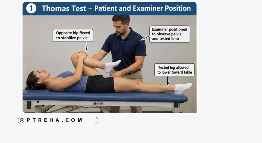
The examiner usually stands beside the patient where the pelvis, tested thigh, and opposite hip can be observed simultaneously.
The examiner should:
- Monitor pelvic position.
- Observe the lumbar spine.
- Control or assist the flexed hip when necessary.
- Observe movement of the tested thigh.
- Avoid forcing the tested hip downward.
- Compare sides when clinically appropriate.
- Note pain, guarding, and compensatory movement.
The test should be performed slowly enough that compensations can be identified.
How to Perform the Thomas Test
A commonly used classic Thomas Test protocol can be performed as follows:
- Position the patient supine on the examination table.
- Observe the resting position of the pelvis and lumbar spine.
- Flex both hips toward the trunk enough to reduce excessive lumbar lordosis and establish a controlled pelvic starting position.
- Ask the patient to hold one knee toward the chest, or assist them in maintaining this position.
- Allow the opposite leg—the test side—to lower toward the examination table.
- Observe the relationship between the posterior thigh and the table.
- Monitor whether the pelvis begins to anteriorly rotate as the leg lowers.
- Observe the lumbar spine for compensatory extension.
- Ask whether the maneuver produces discomfort.
- Repeat on the opposite side if comparison is clinically useful.
The movement should remain comfortable and controlled.
Pushing the thigh toward the table can change the nature of the test and may provoke symptoms that were not present during normal passive positioning.
What Constitutes a Positive Thomas Test?
The terms positive and negative Thomas Test are commonly used in clinical examination, particularly when discussing hip flexion contracture.
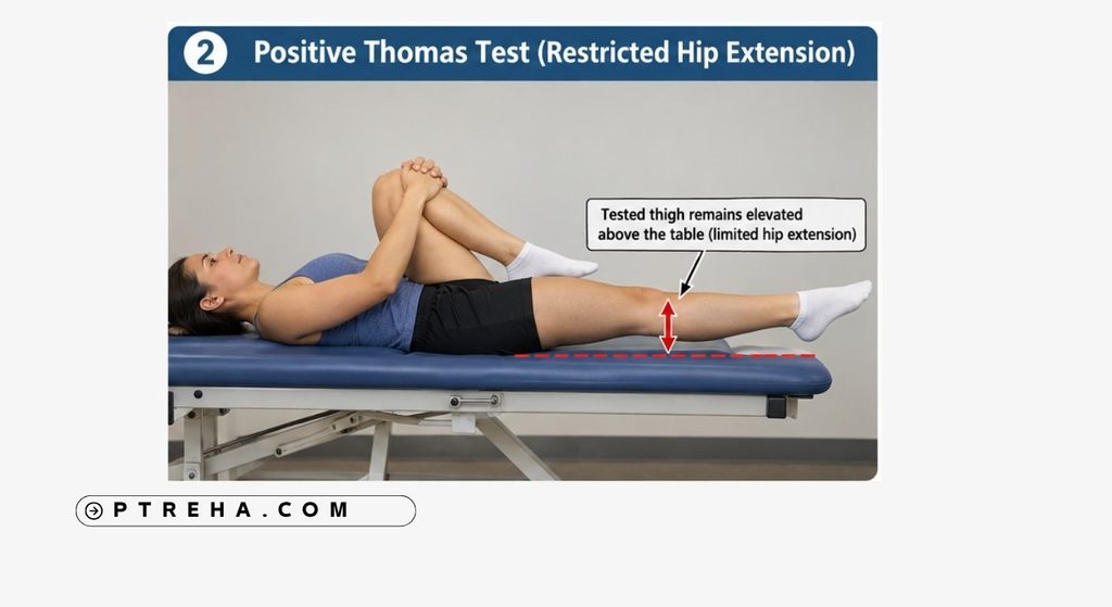
A finding commonly described as positive occurs when the tested thigh cannot lower toward the examination surface without loss of the controlled pelvic position.
In other words, when the contralateral hip is held in sufficient flexion to stabilize the pelvis, the tested hip remains flexed rather than extending toward the examination table.
However, the clinician should interpret this as an examination finding consistent with restricted hip extension, rather than proof of a particular tissue diagnosis.
The test does not independently determine whether the restriction comes from:
- Iliopsoas-related stiffness.
- Other anterior soft tissues.
- Joint or capsular restriction.
- Pain-related guarding.
- Altered pelvic mechanics.
- Another musculoskeletal factor.
That distinction is clinically important.
What Does a Positive Thomas Test Mean?
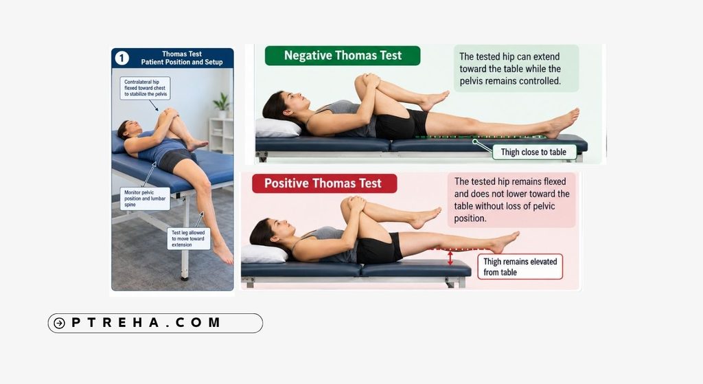
A positive Thomas Test suggests that the tested hip demonstrates a limitation in extension under the conditions of the examination.
The finding may be consistent with hip flexion contracture or restricted hip extension, depending on the patient’s clinical presentation and the protocol used.
It does not establish a specific diagnosis.
For example, it would be inappropriate to conclude from the Thomas Test alone that a patient has an isolated iliopsoas contracture.
Several structures cross the anterior hip, and joint mobility, pelvic positioning, pain, and muscle guarding can all affect the observed movement.
Interpretation should therefore be combined with:
- Passive hip range-of-motion assessment.
- Active movement testing.
- Symptom behavior.
- Palpation when appropriate.
- Functional movement assessment.
- Gait analysis when relevant.
- Other findings from the hip and lumbar examination.
“A restricted Thomas Test is a finding to explain—not a diagnosis to label.”
Negative Thomas Test
A clinically negative Thomas Test generally describes a situation in which the tested hip can move toward the examination surface while pelvic position remains appropriately controlled.
This suggests that an obvious hip flexion contracture was not demonstrated under that testing protocol.
It does not prove that all hip-flexor structures have normal extensibility.
It also does not exclude:
- Hip pathology.
- Exercise-related mobility problems.
- Dynamic movement limitations.
- Pain occurring during other activities.
- Mild restrictions not reproduced by the test.
- Compensations occurring during walking or sport.
Clinical examination should therefore not stop because the Thomas Test appears negative.
Classic Thomas Test vs Modified Thomas Test
The two terms are sometimes used interchangeably, but they should be distinguished.
Classic Thomas Test
The classic test focuses primarily on whether the tested hip can extend adequately while the opposite hip is maintained in flexion and the pelvis is controlled.
It is particularly associated with assessment of hip flexion contracture.
Modified Thomas Test
The Modified Thomas Test generally positions the patient near the end of the examination table, allowing the tested limb to hang more freely.
Clinicians may observe:
- Hip position.
- Knee position.
- Thigh orientation.
- Pelvic position.
The modified test has frequently been used to estimate flexibility involving the hip-flexor region and rectus femoris.
However, clinicians should be careful about assigning an abnormal finding to one isolated muscle based only on limb position.
Research has also demonstrated that pelvic control materially affects Modified Thomas Test measurements.
One validation study in healthy college students found that uncontrolled pelvic tilt substantially influenced measured hip-extension position, reinforcing the importance of standardized pelvic positioning.
This is one reason different Thomas Test protocols should not be treated as interchangeable measurements.
Anatomy Involved in the Thomas Test
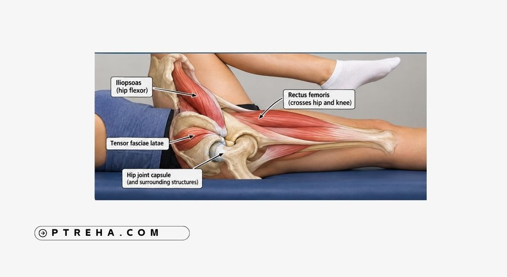
The Thomas Test involves several anatomical regions rather than a single isolated muscle.
Relevant structures include:
- Hip joint.
- Pelvis.
- Lumbar spine.
- Iliopsoas muscle group.
- Rectus femoris.
- Tensor fasciae latae.
- Other anterior hip soft tissues.
- Hip joint capsule and surrounding connective tissues.
Iliopsoas
The iliopsoas contributes to hip flexion and crosses the anterior region of the hip.
Restriction involving this region may influence hip extension, but the Thomas Test does not selectively isolate iliopsoas tissue.
Rectus femoris
The rectus femoris crosses both the hip and knee.
Because knee position changes its length, findings from Modified Thomas Test protocols can be influenced by the knee position as well as the hip.
Pelvis and lumbar spine
These regions are particularly important.
Anterior pelvic rotation can create the appearance of greater hip extension, while lumbar extension may compensate for limited motion at the hip.
For this reason, watching only the thigh is insufficient.
Conditions and Findings Associated With the Thomas Test
The Thomas Test may contribute to assessment of patients with:
- Hip flexion contracture.
- Restricted hip extension.
- Lower-quarter mobility limitations.
- Altered pelvic movement.
- Reduced extension during gait.
- Mobility limitations following prolonged inactivity.
- Selected neurological or orthopedic conditions associated with contracture.
- Rehabilitation-related hip mobility limitations.
These are associations and examination applications, not conditions that the Thomas Test independently diagnoses.
For example, a patient with altered gait may demonstrate reduced hip extension and a restricted Thomas Test, but the test alone cannot establish that the hip restriction caused the gait pattern.
Clinical Significance
The main clinical value of the Thomas Test is contextual.
A therapist can use the finding to help answer questions such as:
- Does the hip appear capable of extending adequately?
- Does the pelvis compensate when the thigh moves toward extension?
- Is there meaningful asymmetry?
- Does the finding correspond with limitations observed during walking?
- Does the test reproduce symptoms?
- Does hip mobility change during rehabilitation?
Suppose a patient demonstrates limited trailing-limb position during gait.
If examination also reveals restricted passive hip extension and a comparable restriction during the Thomas Test, the findings together may strengthen the case that hip mobility deserves attention during rehabilitation.
That still does not establish that the Thomas Test result is the sole cause of the gait change.
Clinical reasoning should integrate the entire examination.
Reliability and Measurement Considerations
Reliability depends considerably on how the test is performed and measured.
A recent systematic review and meta-analysis examining clinical tests for hip-flexor flexibility found substantial methodological variability, particularly for the Thomas Test and Modified Thomas Test.
The authors specifically emphasized the need for more standardized protocols rather than treating reliability as a fixed property of the test.
This has practical implications.
Clinicians monitoring a patient over time should try to reproduce:
- Patient position.
- Pelvic stabilization.
- Examiner technique.
- Table position.
- Knee position.
- Measurement method.
- Side tested first.
- Instructions given to the patient.
A change between visits becomes more difficult to interpret when the protocol also changes.
Pelvic stabilization deserves particular attention
Pelvic motion is one of the main potential sources of measurement error.
When the pelvis anteriorly rotates, the femur may appear to extend farther relative to the examination table even though part of that movement occurred through the pelvis and lumbar region.
Therefore, recording a precise angle without defining pelvic position can give the measurement a level of precision that the procedure may not actually support.
Sensitivity and Specificity
Sensitivity and specificity are most appropriate when a test is being evaluated against a defined reference standard for identifying a specified condition.
The Thomas Test is primarily used clinically as an assessment of hip extension restriction or hip flexion contracture, rather than as a stand-alone diagnostic test for a specific disease.
Evidence for hip-flexor flexibility testing also varies according to protocol, population, examiner technique, pelvic stabilization, and the definition of the target finding.
For these reasons, a single sensitivity or specificity value should not be presented as universal diagnostic accuracy for the Thomas Test.
Its result is better interpreted as part of a broader musculoskeletal examination.
Diagnostic Accuracy
The Thomas Test should not be regarded as independently diagnostic of:
- Iliopsoas pathology.
- Rectus femoris pathology.
- Hip impingement.
- Labral injury.
- Hip osteoarthritis.
- Lumbar disorders.
- A specific source of anterior hip pain.
Although Thomas-type maneuvers have been investigated in several clinical contexts, diagnostic claims depend on exactly which maneuver, outcome, reference standard, and population were studied.
For routine physical therapy use, its strongest role is as a clinical examination finding related to hip extension mobility, not as a definitive diagnostic procedure.
Common Mistakes in the Thomas Test
Several technique errors can reduce the clinical usefulness of the test.
Failing to control the pelvis
This is one of the most important errors.
If anterior pelvic tilt occurs while the leg lowers, the examiner may overestimate available hip extension.
Watching only the thigh
The pelvis and lumbar spine should be observed at the same time.
Forcing the leg toward the table
The test is an examination maneuver, not an aggressive stretch.
Force can provoke pain and change the result.
Automatically blaming the iliopsoas
A restricted position does not prove isolated iliopsoas tightness.
Mixing classic and modified protocols
Measurements obtained from different protocols should not automatically be compared as though they represent the same procedure.
Ignoring symptoms
Pain, apprehension, or guarding can restrict motion independently of tissue extensibility.
Ignoring knee position
This is particularly relevant during Modified Thomas Test variations because rectus femoris crosses both the hip and knee.
Using arbitrary universal cutoffs
A single angle should not automatically be labeled normal or abnormal without considering the measurement protocol, patient population, and available reference data.
Failing to document the technique
If the test is being repeated during rehabilitation, the protocol should be documented clearly enough to reproduce it.
Tips for Performing the Thomas Test
For a more clinically useful examination:
- Establish pelvic position before lowering the test leg.
- Observe the lumbar spine throughout the maneuver.
- Perform the movement slowly.
- Avoid pushing the thigh toward the table.
- Compare sides when comparison is meaningful.
- Document whether pain occurred.
- Record compensatory pelvic movement.
- Specify whether the classic or Modified Thomas Test was used.
- Use the same protocol during follow-up testing.
- Interpret the test alongside other hip examination findings.
When quantitative measurement is required, clinicians should use a defined protocol rather than casually estimating an angle from the table.
Consistency often matters more than creating an artificial appearance of precision.
How the Thomas Test Fits Into a Physical Therapy Examination
The Thomas Test becomes more valuable when its finding matches other clinically relevant information.
For example, consider a patient who demonstrates limited hip extension during walking.
A useful examination may include:
- Observation of gait.
- Active hip motion.
- Passive hip motion.
- Thomas Test.
- Pelvic and lumbar movement assessment.
- Relevant strength testing.
- Functional tasks.
- Symptom reproduction.
- Patient-specific activity limitations.
If several findings point toward restricted hip extension, the therapist can determine whether mobility-focused intervention is appropriate.
Conversely, if the Thomas Test appears restricted but gait and functional activities are unaffected, the finding may have less clinical importance.
Treatment should target meaningful impairments related to the patient’s function, rather than attempting to normalize every examination finding.
Summary
- The Thomas Test is a clinical musculoskeletal assessment primarily used to evaluate hip extension restriction or hip flexion contracture.
- The opposite hip is flexed while the tested limb moves toward extension, with careful attention to pelvic and lumbar control.
- Failure of the tested thigh to move appropriately toward the table can be interpreted as a finding consistent with restricted hip extension, but it does not identify a specific tissue diagnosis.
- Pelvic positioning and protocol standardization can substantially influence the result, especially when quantitative measurements are used.
- The Thomas Test should be interpreted alongside range of motion, symptoms, gait, functional movement, and the rest of the physical therapy examination.
The Thomas Test remains a practical component of the hip examination when performed carefully.
Its greatest value comes from standardized technique and thoughtful integration with the patient’s overall clinical presentation rather than interpreting one position of the thigh as a diagnosis.