
The Posterior Sag Sign is a passive orthopedic special test used to observe whether the tibia rests farther posteriorly than expected relative to the femur. It can support suspicion of posterior cruciate ligament (PCL) insufficiency, but it does not establish the diagnosis by itself.
What Is the Posterior Sag Sign?
The test compares the resting anterior step-off of the tibia with the knee flexed. Loss of the usual step-off, or visible posterior sag compared with the other knee, is an abnormal finding that should be interpreted with the history, other examination findings, and imaging when indicated.
Purpose and Indications
The Posterior Sag Sign is used when the history or symptoms raise concern about posterior tibial instability.
- Assess the resting relationship between the tibia and femur.
- Screen for findings consistent with PCL injury or chronic PCL insufficiency.
- Compare sides when the opposite knee is suitable as a reference.
- Guide the need for a fuller ligament examination or referral.
Contraindications and Precautions
The maneuver is observational and does not require a forceful stress, but positioning may be inappropriate in an acutely injured knee.
- Defer or modify the test when fracture, dislocation, or another injury needing urgent stabilization is suspected.
- Avoid forcing knee or hip flexion when motion is severely painful or restricted.
- Support the limb if muscle guarding prevents a relaxed resting position.
- Use caution after recent surgery and follow the treating team’s precautions.
Patient and Examiner Positions
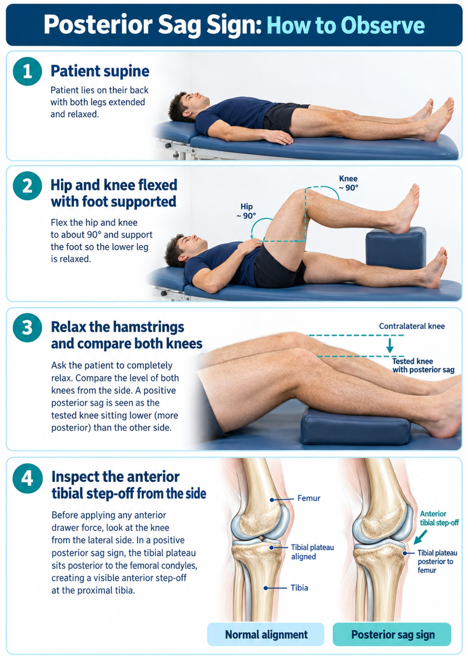
- Patient: supine with the hip and knee flexed and the foot supported so the leg can relax.
- Comparison: both knees may be positioned together when the opposite side is appropriate for comparison.
- Examiner: positioned to view the anterior tibial plateau and tibial tuberosity from the side.
- Key requirement: the hamstrings should be relaxed because guarding can alter the apparent tibial position.
How to Perform the Posterior Sag Sign
- Explain the examination and obtain consent.
- Position the patient supine with the hip and knee flexed, commonly near a right angle, without forcing the range.
- Support the foot and ask the patient to relax the leg completely.
- View the knee from the side and compare the anterior step-off of the tibia relative to the femur.
- Compare with the other knee when appropriate and document pain, guarding, swelling, and asymmetry.
Test Findings and Interpretation
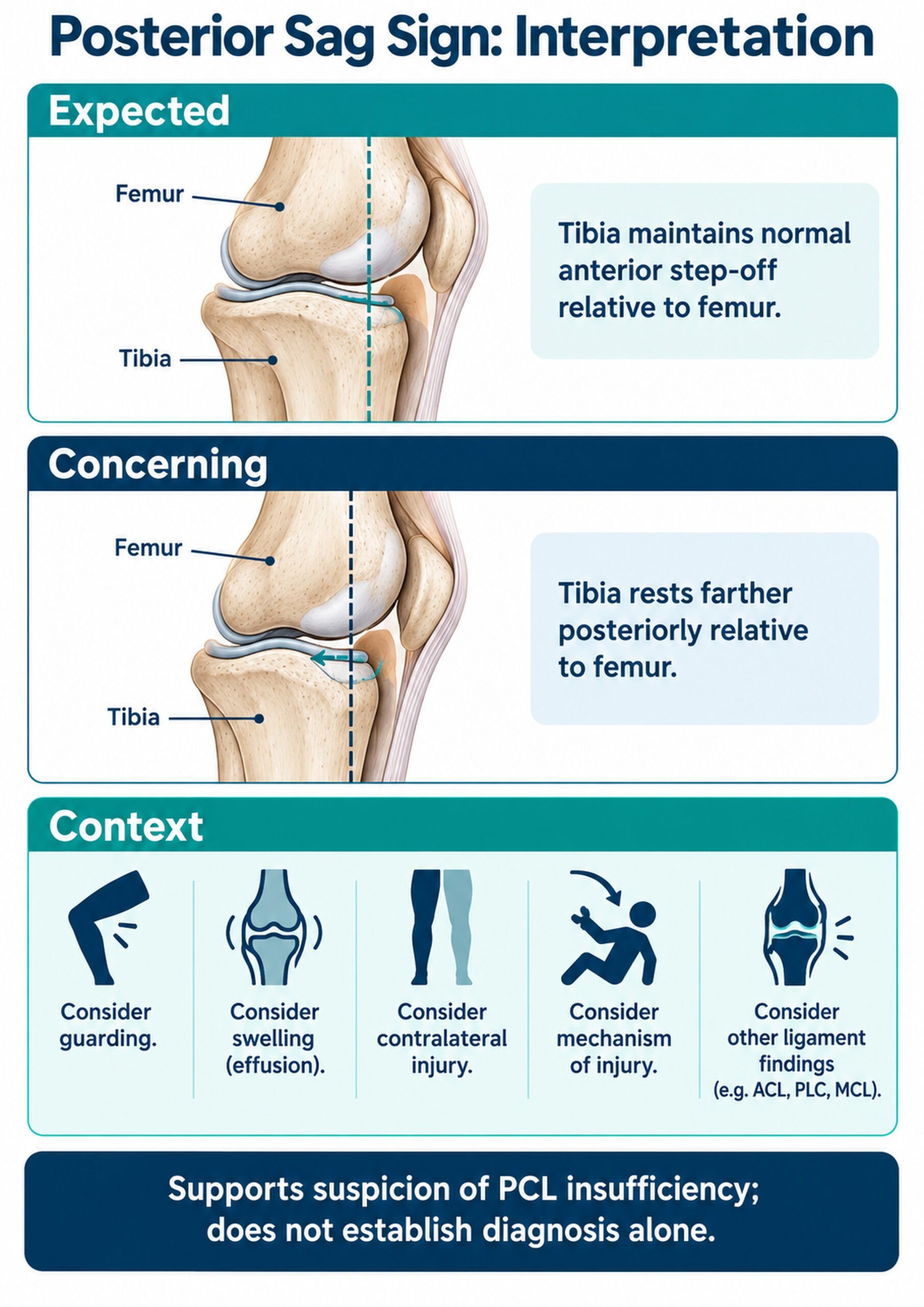
An abnormal Posterior Sag Sign is visible posterior resting displacement of the tibia, with reduced or reversed anterior tibial step-off compared with expected alignment or the other knee. A normal-appearing step-off reduces concern from this observation alone but does not exclude a PCL injury.
- Record whether the finding is present, absent, or difficult to interpret.
- Describe the comparison side rather than relying only on “positive” or “negative.”
- Note whether pain, effusion, guarding, or limited flexion affected the test.
What an Abnormal Finding Means
Posterior sag is associated with reduced restraint to posterior tibial translation and can support a PCL injury hypothesis. It does not identify injury grade, chronicity, associated ligament injury, or treatment needs on its own.
A current review of PCL diagnostic standards emphasizes combining the history, multiple examination maneuvers, and appropriate imaging rather than relying on a single test.
Relevant Anatomy
- The PCL runs from the posterior tibia toward the medial femoral condyle.
- It is a major restraint to posterior translation of the tibia relative to the femur.
- The joint capsule, menisci, collateral ligaments, posterolateral structures, and muscles also influence knee stability.
- The test observes joint alignment; it does not isolate one structure.
Conditions Associated With Posterior Sag
- Isolated PCL injury
- Combined ligament injury involving the PCL
- Chronic posterior knee instability
- Traumatic patterns such as a force to the proximal tibia with the knee flexed
These are associations and clinical possibilities, not diagnoses produced by the sign.
Clinical Significance
- Adds a passive resting-alignment observation to the ligament examination.
- Helps determine whether posterior instability needs further testing.
- Provides a baseline description for follow-up when the same position and method are used.
- May influence decisions about imaging, specialist referral, bracing, and rehabilitation precautions.
Evidence and Diagnostic Accuracy
Reliable universal sensitivity and specificity values for the Posterior Sag Sign are not established across populations and protocols. A systematic review of the physical examination for PCL injury found that diagnostic accuracy was largely unknown, so a single observation should not be used as a stand-alone rule-in or rule-out test.
A broader review of PCL evaluation and management describes examination within a comprehensive assessment that includes mechanism, posterior drawer testing, associated ligament assessment, and imaging as clinically indicated.
Common Mistakes and Practical Tips
- Viewing from above: inspect from the side to judge the anterior step-off.
- Active hamstrings: pause, support the foot, and cue relaxation.
- Unequal positions: match hip and knee flexion before comparing sides.
- Ignoring swelling: document effusion because it can obscure landmarks and limit flexion.
- Calling it diagnostic: integrate the finding with the full examination.
Limitations
Body habitus, swelling, pain, guarding, bilateral laxity, prior injury, and variation in positioning can affect visibility and comparison. The sign does not quantify translation or determine which associated structures are injured.
Summary
- The Posterior Sag Sign observes passive posterior resting displacement of the tibia.
- The patient must be supported and relaxed, with the knee viewed from the side.
- Reduced anterior tibial step-off supports suspicion of PCL insufficiency.
- The finding requires confirmation within a comprehensive examination and, when indicated, imaging.
- Universal diagnostic-accuracy values are not established for all settings.
Used carefully, the Posterior Sag Sign is a useful part of knee ligament assessment, but its value comes from integration with the full clinical picture.