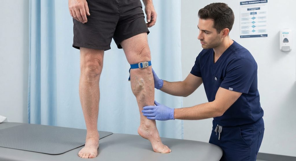
The Perthes Test is a traditional clinical venous assessment used in patients with lower-limb varicosities to obtain information about the function of the deep venous system and the relationship between superficial, perforating, and deep veins. It is performed while the patient is standing and activating the calf-muscle pump, with a tourniquet used to alter superficial venous flow. The test is best understood as a clinical assessment of venous drainage rather than a stand-alone diagnostic test.
What Is the Perthes Test?
The Perthes Test is a clinical venous function test, rather than an orthopedic or functional performance test. Its traditional purpose is to assess whether blood from superficial varicose veins can drain effectively through the deep venous system when the superficial venous pathway is partially occluded.
The examination uses muscular activity—commonly walking or repeated heel raises—to activate the calf-muscle pump. Changes in the appearance and distension of superficial varicosities are then observed.
Different descriptions of the Perthes Test use different tourniquet locations and activity protocols. Therefore, the examiner should identify the protocol being used rather than treating one variation as the only standardized version.
Importantly, the Perthes Test should not be confused with Legg-Calvé-Perthes disease, which is an entirely different pediatric hip condition.
Purpose of the Perthes Test
The traditional purposes of the Perthes Test include:
- Assessing the functional adequacy of deep venous drainage in a limb with varicosities.
- Observing whether superficial varicosities decompress during activation of the calf-muscle pump.
- Assessing the interaction between the superficial and deep venous systems.
- Providing clinical information that may contribute to the assessment of chronic venous disease.
- Historically helping clinicians consider whether superficial varicosities might be associated with impaired deep venous drainage before treatment.
The test does not directly visualize venous anatomy and should not be regarded as a substitute for duplex ultrasonography when detailed assessment of venous patency or reflux is required. Modern clinical evaluation commonly relies on ultrasound-based assessment when definitive anatomical information is needed.
Perthes Test Indications
The Perthes Test may be considered during clinical assessment of:
- Visible lower-limb varicose veins.
- Suspected chronic venous disease.
- Patients in whom the clinician wants bedside information about deep venous drainage.
- Historical or teaching examinations of superficial and deep venous relationships.
- Clinical assessment performed before consideration of treatment for varicose veins, when appropriate.
Its usefulness depends on the clinical context and the protocol used. A finding should always be interpreted alongside the history, physical examination, and, when indicated, vascular imaging.
Perthes Test Contraindications and Precautions
A universal list of test-specific contraindications is not consistently established in the clinical literature. The main issue is patient safety and appropriate clinical selection.
Relevant precautions include:
- Do not force walking, heel raises, or other activity in a patient who cannot safely perform the movement.
- Stop the examination if the patient develops significant pain, dizziness, marked discomfort, or other concerning symptoms.
- Use caution in patients with acute lower-limb symptoms in whom a vascular emergency or other serious pathology is suspected.
- Avoid excessive tourniquet pressure or prolonged compression.
- Consider the patient’s mobility, balance, skin condition, and ability to tolerate standing.
- If acute deep-vein thrombosis is clinically suspected, do not use a bedside maneuver as a substitute for appropriate medical evaluation and diagnostic imaging.
The Perthes Test is an observational clinical test. It should not delay investigation when the clinical presentation raises concern for acute vascular disease.
Patient Position
The commonly described position is:
- The patient stands upright with the affected lower limb visible to the examiner.
- Varicose veins are identified while the patient is standing.
- A tourniquet is applied at a selected level according to the protocol being used.
- The patient is then asked to activate the calf-muscle pump, commonly by walking or performing repeated heel raises.
Traditional descriptions differ in whether the tourniquet is placed below the knee, around the thigh, or at another level selected to occlude a superficial venous pathway.
Examiner Position
The examiner should:
- Stand beside the patient in a position that allows continuous observation of the varicosities.
- Apply the tourniquet at the level specified by the chosen protocol.
- Observe the superficial veins before, during, and after muscular activity.
- Monitor the patient for pain, instability, or other symptoms.
- Compare findings with the opposite limb when clinically appropriate, while recognizing that bilateral venous disease may occur.
Good lighting and adequate exposure of the lower limb are important because the test depends substantially on visual observation.
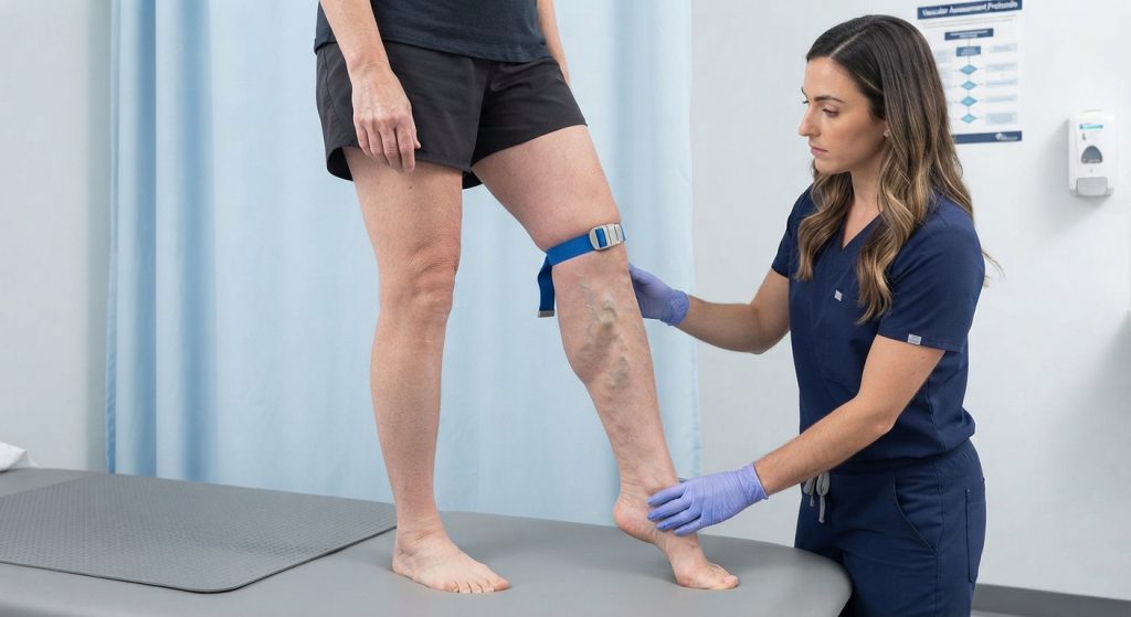
How to Perform the Perthes Test
One commonly described clinical approach is as follows:
- Position the patient standing and inspect the superficial varicosities.
- Select the tourniquet level according to the protocol being used.
- Apply the tourniquet firmly enough to alter superficial venous flow without causing excessive compression.
- Ask the patient to activate the calf-muscle pump. Depending on the protocol, this may involve walking or repeated heel raises.
- Observe the varicosities during the maneuver.
- Note whether the superficial veins become less distended, remain distended, or become more prominent.
- Stop the examination if the patient develops significant symptoms or cannot safely continue.
- Interpret the observation in the context of the selected protocol and the remainder of the vascular examination.
The purpose of muscular activity is to promote venous return through the calf-muscle pump. If the deep venous pathway is functioning adequately, blood can be redirected through the available venous channels and superficial varicosities may decompress.
Interpretation of Perthes Test Findings
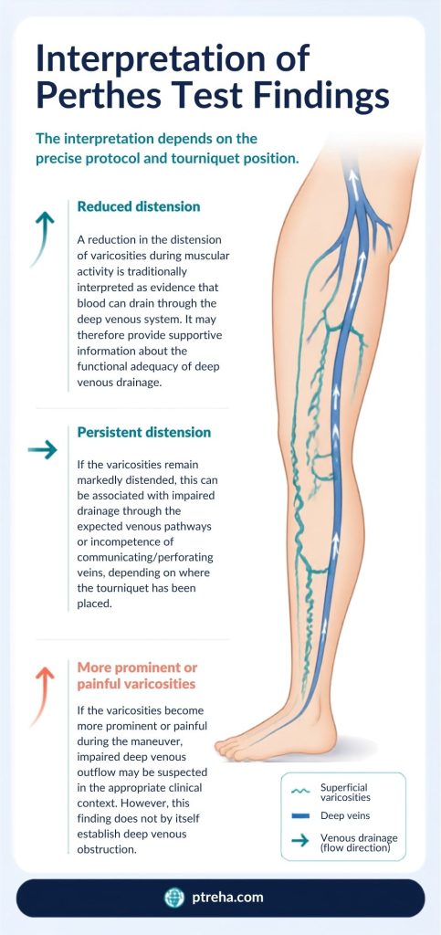
The interpretation depends on the precise protocol and tourniquet position.
A reduction in the distension of varicosities during muscular activity is traditionally interpreted as evidence that blood can drain through the deep venous system. It may therefore provide supportive information about the functional adequacy of deep venous drainage.
If the varicosities remain markedly distended, this can be associated with impaired drainage through the expected venous pathways or incompetence of communicating/perforating veins, depending on where the tourniquet has been placed.
If the varicosities become more prominent or painful during the maneuver, impaired deep venous outflow may be suspected in the appropriate clinical context. However, this finding does not by itself establish deep venous obstruction.
“The Perthes Test is best interpreted as a venous-flow assessment, not as a stand-alone diagnosis.”
The terminology of “positive” and “negative” Perthes Test is used in some clinical sources, but interpretation is more useful when the actual observed venous response is documented.
What Does an Abnormal Perthes Test Mean?
An abnormal response may indicate that superficial venous blood is not being effectively redirected through the expected deep venous pathways during muscle-pump activity.
Possible explanations depend on the test protocol and may include:
- Impaired deep venous drainage.
- Deep venous obstruction.
- Dysfunction of communicating or perforating veins.
- Complex venous reflux involving more than one venous pathway.
The finding should not be interpreted as proof of a particular lesion or diagnosis.
A bedside Perthes finding also cannot reliably determine the exact anatomical site or cause of venous dysfunction. Duplex ultrasound provides substantially more anatomical and hemodynamic information when detailed assessment is required.
Anatomy Involved in Perthes Test
The Perthes Test involves the functional interaction of several components of the lower-limb venous system:
- Superficial veins — including the saphenous venous pathways and their tributaries, which can become visibly dilated in varicose disease.
- Deep veins — veins located within the deep compartments of the limb that provide an important route for venous return.
- Perforating or communicating veins — vessels connecting superficial and deep venous systems.
- Venous valves — structures that help regulate the direction of venous blood flow.
- Calf-muscle pump — muscular contraction compresses veins and contributes to venous return from the lower limb.
The test does not isolate any single vein, valve, or muscle. Its observation reflects the behavior of an interconnected venous system.
Conditions Detected / Associated With the Test
The Perthes Test has traditionally been associated with clinical assessment of:
- Lower-limb varicose veins.
- Chronic venous insufficiency.
- Abnormal superficial-to-deep venous communication.
- Possible impairment of deep venous outflow.
- Venous obstruction in an appropriate clinical setting.
The term “associated with” is important. The Perthes Test alone does not establish the presence, severity, anatomical location, or cause of chronic venous disease.
Modern evaluation of venous disease generally incorporates clinical examination and, when appropriate, duplex ultrasound rather than relying on traditional bedside tests alone.
Clinical Significance
The Perthes Test can still be useful from a clinical-education and bedside-assessment perspective because it demonstrates an important physiological concept: lower-limb venous return depends on coordinated function of the superficial veins, perforating connections, deep veins, valves, and calf-muscle pump.
Clinically, the observation may help the examiner:
- Recognize abnormal venous filling or emptying.
- Understand the relationship between superficial and deep venous drainage.
- Decide whether further vascular assessment is warranted.
- Document changes in visible varicosities during muscle-pump activation.
- Recognize situations in which a simple bedside examination is insufficient.
Traditional bedside venous tests have limitations, and modern ultrasound techniques have largely replaced them when precise assessment of venous reflux or patency is required.
Sensitivity and Specificity
Published clinical literature has described diagnostic-performance characteristics for traditional venous examination maneuvers, including the Perthes Test, but these results are dependent on the population, reference standard, protocol, and definition of the clinical finding.
A universal sensitivity or specificity value for the Perthes Test should therefore not be presented as applicable to all patients or protocols.
The test should be considered a supplementary clinical assessment rather than a definitive diagnostic procedure.
Diagnostic Accuracy
The Perthes Test should not be confused with duplex ultrasonography or venography.
Its clinical finding can provide information about venous function, but it does not directly visualize thrombus, reflux, stenosis, or obstruction. Consequently, an abnormal clinical response requires appropriate clinical correlation rather than automatic conversion into a specific vascular diagnosis.
Current clinical resources emphasize ultrasound-based assessment when reliable anatomical and hemodynamic information about the venous system is required.
Common Mistakes in Perthes Test
Common problems include:
- Treating one described protocol as universally standardized.
- Failing to identify the tourniquet level before interpreting the finding.
- Applying excessive tourniquet pressure.
- Observing the veins only before and after exercise instead of throughout the maneuver.
- Asking a patient with poor balance to perform heel raises without adequate safety support.
- Interpreting persistent varicosity as definitive proof of deep-vein obstruction.
- Assuming that the test identifies the precise incompetent vein.
- Using the Perthes Test instead of appropriate vascular imaging when imaging is clinically indicated.
- Confusing the Perthes Test with tests for arterial circulation.
- Confusing the venous Perthes Test with Legg-Calvé-Perthes disease of the hip.
Tips for Performing the Perthes Test
For a useful clinical examination:
- Clearly document the tourniquet location and activity protocol.
- Examine the patient standing so that superficial venous distension can be assessed.
- Ensure the patient can safely walk or perform the selected movement.
- Watch for both venous decompression and increasing distension.
- Ask about pain rather than assuming that discomfort has a specific vascular cause.
- Interpret the result together with inspection, palpation, medical history, and other vascular findings.
- Use duplex ultrasound when the clinical question requires anatomical localization or confirmation of venous pathology.
- Avoid describing the result simply as “positive” without explaining what actually happened to the varicosities.
Summary
- The Perthes Test is a traditional clinical venous function assessment, not an orthopedic or neurological special test.
- It examines the behavior of superficial varicosities while the calf-muscle pump is activated and superficial venous flow is modified by a tourniquet.
- Reduction in varicosity distension generally supports adequate drainage through available venous pathways, while persistent or increased distension may indicate abnormal venous drainage depending on the protocol.
- The result does not independently diagnose deep-vein obstruction, chronic venous insufficiency, or a specific incompetent vein.
- Protocols differ, particularly regarding tourniquet position and the method of muscle-pump activation, so the protocol should always be documented.
- Duplex ultrasound is preferred when detailed assessment of venous patency, reflux, or anatomy is clinically required.
The Perthes Test remains useful for understanding lower-limb venous physiology and for structured bedside assessment, but its findings should be interpreted cautiously. In contemporary practice, it is best used as one component of a broader vascular examination rather than as a definitive diagnostic test.
External Links and Resources
- Encyclopedia
- Journal of Dermatology
- PMC — Clinical assessment of chronic venous insufficiency and traditional venous tests.
- American Family Physician — Clinical assessment and management of varicose veins.
- Thieme / Via Medici — Clinical examination of the vascular system and limitations of traditional venous function tests.
- Journal of Dermatology — Earlier guideline discussion of the Perthes Test.