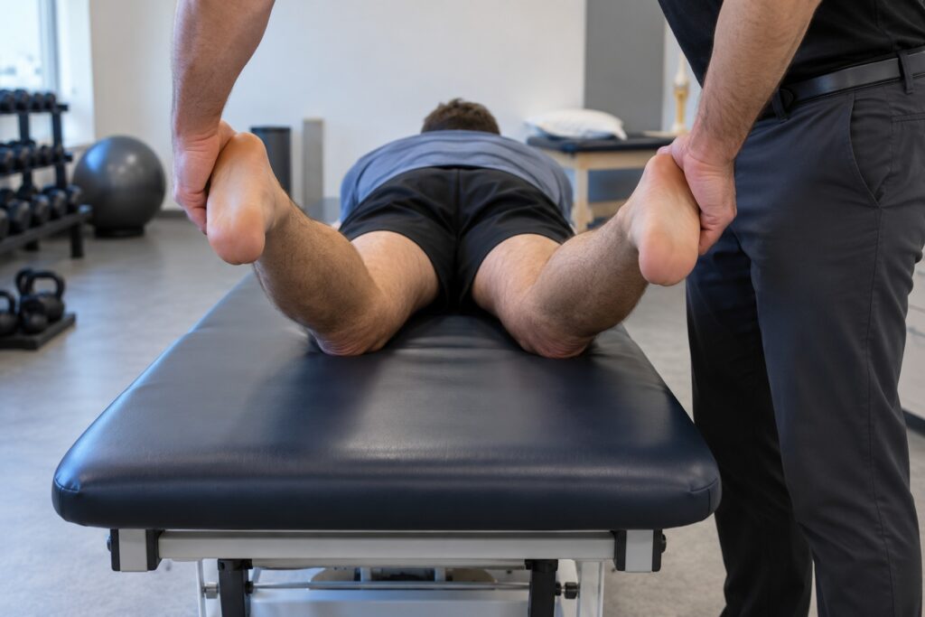
The Dial Test is an orthopedic special test that compares passive external rotation of the lower legs at different knee-flexion angles. It can contribute to assessment of posterolateral and cruciate ligament injury patterns, but overlapping findings mean it should never be interpreted alone.
What Is the Dial Test?
The test observes and compares the thigh-foot angle while equal external-rotation torque is applied to both feet. The common prone protocol examines the knees at 30° and 90° of flexion, with side-to-side asymmetry recorded as a finding rather than treated as a diagnosis.
Purpose and Indications
- Assess side-to-side differences in passive external tibial rotation.
- Investigate suspected posterolateral rotatory instability after knee trauma.
- Contribute to examination of possible posterolateral corner (PLC), PCL, or combined ligament injury.
- Help plan further testing or referral when instability symptoms are present.
Contraindications and Precautions
The Dial Test should not be forced through a painful or unstable range.
- Defer the maneuver when fracture, dislocation, or another injury needing urgent stabilization is suspected.
- Modify or stop when acute pain, swelling, or guarding prevents a relaxed comparison.
- Follow procedure-specific precautions after recent surgery.
- Support the limb and use gentle, matched torque on both sides.
Patient and Examiner Positions
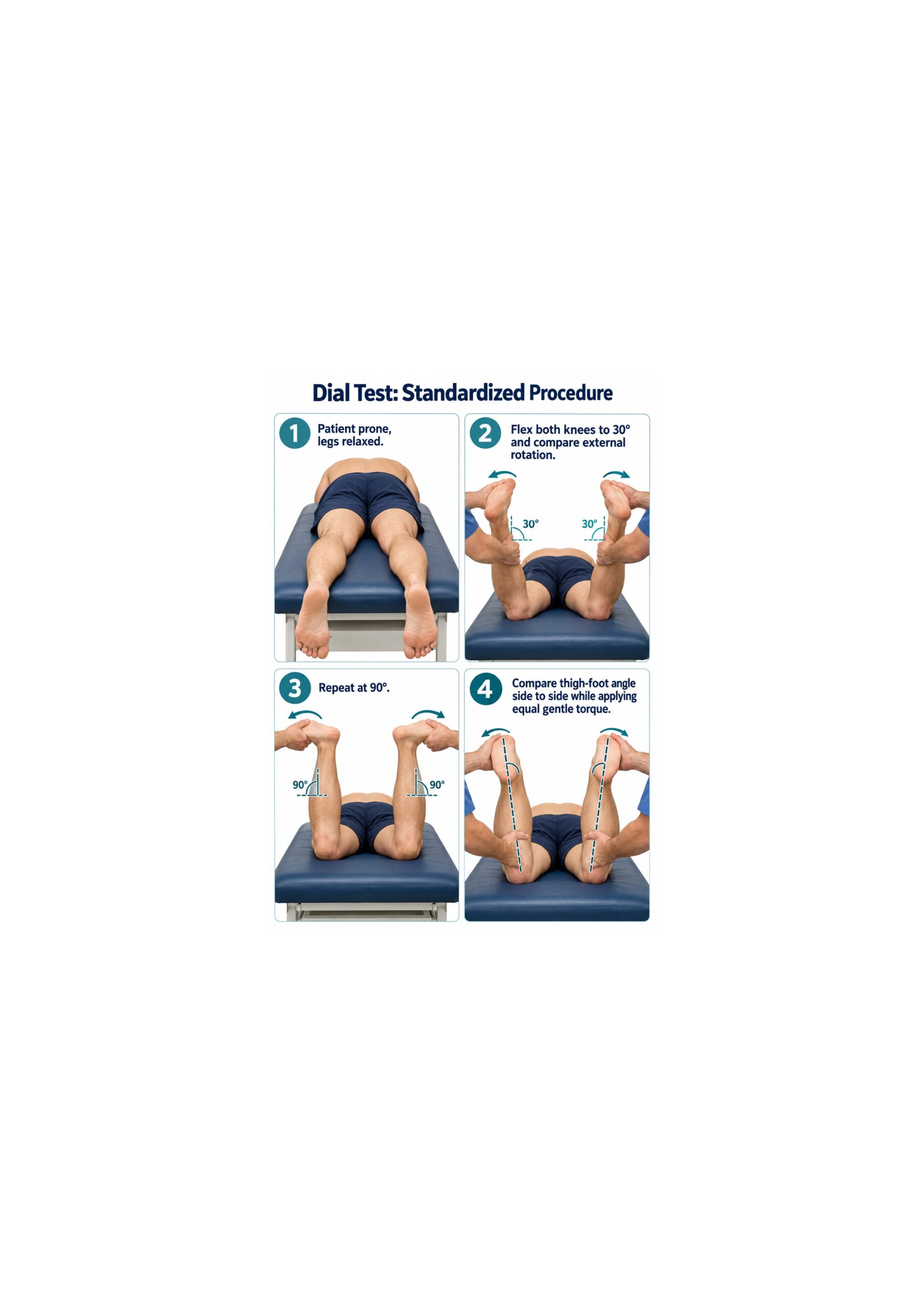
- Patient: prone with the hips neutral and both legs relaxed.
- First position: both knees flexed to 30°.
- Second position: both knees flexed to 90°.
- Examiner: at the foot of the table, holding both heels and maintaining matched knee flexion.
A supine variation is also described, but position should be documented because technique can affect the result.
How to Perform the Dial Test
- Explain the procedure, obtain consent, and inspect both legs for resting asymmetry.
- Position the patient prone and flex both knees to 30°.
- Hold the heels, relax the ankles, and apply gentle equal external-rotation torque.
- Compare the thigh-foot angles and note pain, guarding, and the endpoint.
- Repeat the same comparison at 90° of knee flexion.
- Return the legs to rest and document the position, method, and side-to-side pattern.
Test Findings and Interpretation
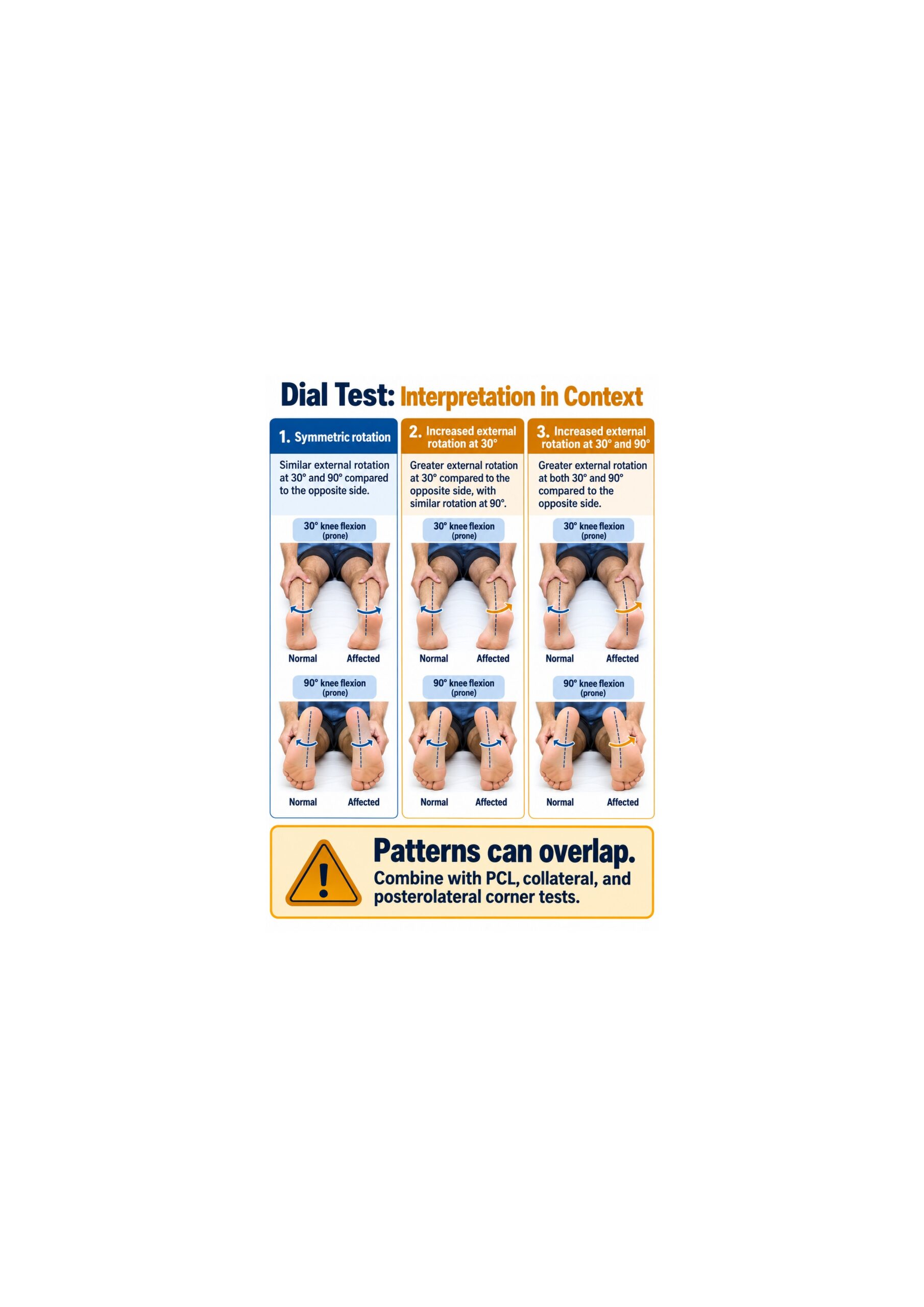
A concerning finding is visibly greater external rotation on one side compared with the other under matched positioning and torque. Traditional interpretation associates asymmetry mainly at 30° with a PLC pattern and asymmetry at both 30° and 90° with combined PLC and PCL involvement.
Those patterns are not specific enough to stand alone. Medial instability, baseline rotational asymmetry, guarding, and technique can produce similar findings.
What an Abnormal Finding Does and Does Not Mean
- It supports further assessment of rotational knee instability.
- It does not identify a single injured structure by itself.
- It does not establish injury grade or determine treatment.
- It should be compared with collateral, cruciate, and other posterolateral tests.
A diagnostic-accuracy study of Dial Test interpretations found that several injury patterns can overlap, supporting cautious use alongside other examination findings.
Relevant Anatomy
- The fibular collateral ligament contributes to varus restraint.
- The popliteus tendon and popliteofibular ligament contribute to posterolateral stability.
- The PCL contributes to posterior and rotational stability, particularly as knee flexion increases.
- Medial structures can also influence apparent external rotation.
The Dial Test loads a system of stabilizers; it does not isolate one ligament.
Conditions Associated With an Abnormal Dial Test
- Posterolateral corner injury
- Combined PLC and PCL injury
- Multiligament knee injury
- Medial collateral or posteromedial instability that mimics a posterolateral pattern
These are possible associations. The finding requires correlation with mechanism, symptoms, other tests, and imaging when indicated.
Clinical Significance
- Adds a rotational comparison to a complete knee ligament examination.
- May raise concern for an injury pattern that needs specialist assessment.
- Helps direct additional varus, valgus, posterior, and posterolateral testing.
- Can be documented over time when the same position and method are used.
Evidence and Diagnostic Accuracy
Diagnostic performance is protocol- and population-dependent, and universal sensitivity or specificity values should not be assigned to the manual test. A clinical study of manual Dial Test reliability reported questionable rater agreement, reinforcing the need for standardized positioning and cautious interpretation.
A comprehensive review of posterolateral corner injuries describes the Dial Test as one component of a broader examination that includes mechanism, varus stress, posterolateral drawer, reverse pivot-shift, and cruciate assessment.
Common Mistakes and Practical Tips
- Unequal torque: rotate both feet gently and symmetrically.
- Changing hip position: keep the pelvis and thighs aligned on the table.
- Measuring only the feet: compare the thigh-foot relationship, not foot posture alone.
- Ignoring guarding: repeat only after the patient relaxes; do not force the range.
- Using one flexion angle: document findings at both protocol positions.
- Calling the test diagnostic: integrate it with the full ligament examination.
Limitations
Natural asymmetry, tibial torsion, foot posture, examiner force, knee-flexion accuracy, pain, and muscle guarding can alter the apparent rotation. Bilateral injury also reduces the value of the opposite side as a reference.
Summary
- The Dial Test compares passive external rotation side to side.
- The common prone protocol uses 30° and 90° of knee flexion.
- Greater rotation may support concern for PLC, PCL, medial, or combined injury patterns.
- Technique and rater agreement limit stand-alone interpretation.
- Use the result with history, other ligament tests, and imaging when indicated.
The Dial Test is most useful as a carefully standardized comparison within a complete knee examination, not as an isolated diagnostic decision.