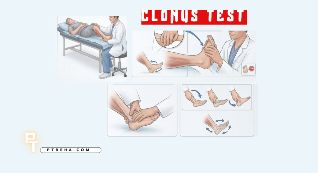
The Clonus Test is a neurological clinical assessment used to identify rhythmic, involuntary muscle contractions produced by a sudden sustained stretch. It is most commonly examined at the ankle and can provide evidence of altered reflex excitability, particularly when considered alongside other upper motor neuron findings such as hyperreflexia, spasticity, weakness, and abnormal plantar responses. The finding should never be interpreted in isolation as a diagnosis.
What Is the Clonus Test?
The Clonus Test is best classified as a neurological clinical assessment of a reflex response, rather than an orthopedic special test, functional performance test, or outcome measure.
Clonus consists of repetitive, rhythmic muscle contractions and relaxations that occur after a muscle is rapidly stretched and the stretch is maintained. At the ankle, the response is commonly elicited by briskly dorsiflexing the foot and maintaining pressure in dorsiflexion.
The test examines the behavior of the stretch-reflex system and the influence of descending motor pathways on spinal reflex activity. A clinically important finding is usually considered in the context of the rest of the neurological examination rather than as an isolated diagnostic result.
“Clonus is a neurological finding, not a diagnosis.”
The ankle is the most commonly examined site, although clonus can also be assessed at other joints, including the patella.
Purpose of the Clonus Test
The assessment may be used to:
- Identify rhythmic involuntary responses produced by a sudden, sustained muscle stretch.
- Look for evidence of increased reflex excitability during a neurological examination.
- Support the assessment of possible upper motor neuron involvement when combined with other examination findings.
- Compare neurological findings between the right and left sides.
- Contribute to the assessment of patients with suspected central nervous system or spinal cord dysfunction.
- Provide additional neurological information when examining changes in tone, reflexes, gait, strength, or coordination.
Clonus is particularly useful when interpreted as part of a broader neurological examination rather than as a stand-alone test.
Clonus Test Indications
Common clinical situations in which clonus may be assessed include:
- Suspected upper motor neuron involvement.
- Hyperreflexia or other abnormal deep tendon reflex findings.
- Suspected spinal cord dysfunction or myelopathy.
- Neurological conditions associated with altered descending motor control.
- Unexplained stiffness, abnormal muscle tone, or changes in gait occurring with other neurological signs.
- Follow-up neurological examination when changes in reflex behavior are clinically relevant.
- Neurological screening or examination after a known central nervous system injury, depending on the clinical context.
The presence of clonus can contribute to clinical reasoning in conditions affecting the brain or spinal cord, but it does not by itself establish the location or cause of a neurological lesion.
Clonus Test Contraindications and Precautions
There is no universally established absolute contraindication specific to the Clonus Test. However, the maneuver should be performed carefully and adapted to the patient’s clinical condition.
Important precautions include:
- Do not use excessive force or unnecessarily aggressive joint movement.
- Consider pain, joint instability, recent surgery, acute injury, or other circumstances in which passive movement may be inappropriate.
- Ensure that the limb is adequately supported and that unnecessary muscle activation is minimized.
- Stop the maneuver if it produces significant pain or the patient cannot tolerate the movement.
- Interpret the finding according to the patient’s age and neurological context rather than applying a universal threshold.
- In patients with acute or evolving neurological symptoms, treat the clonus finding as one component of the overall examination and clinical assessment.
The goal is to elicit the reflex response safely, not to provoke the greatest possible movement.
Patient Position
A commonly used ankle-clonus position includes:
- Position the patient comfortably, commonly in supine.
- Support the lower limb so that the patient can relax as much as possible.
- Allow the knee to remain comfortably positioned rather than requiring a rigid standardized angle.
- Keep the ankle close to a neutral position before the maneuver.
- Ensure that the examiner can control the foot throughout the movement.
The exact positioning can vary between clinical protocols, but relaxation and adequate support are more important than forcing the limb into a particular position.
Examiner Position
The examiner should:
- Stand or sit where the lower limb and foot can be comfortably supported.
- Use one hand to stabilize and support the lower leg when necessary.
- Place the other hand over the forefoot to control ankle movement.
- Maintain sufficient contact to feel the rhythmic response.
- Be positioned so that the movement can be stopped immediately if discomfort occurs.
Good examiner control is essential because clonus depends on the way the stretch is applied and maintained.
How to Perform the Clonus Test
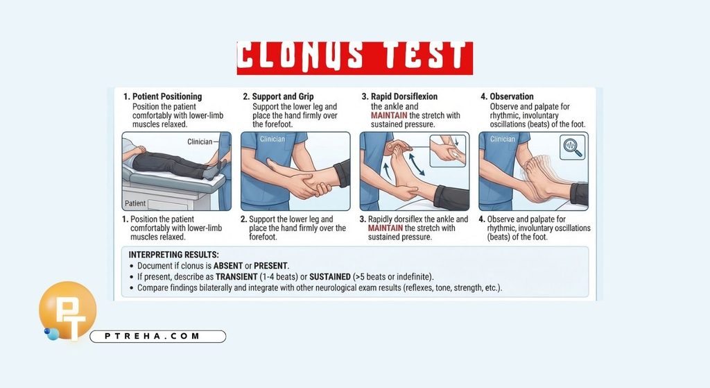
The following describes a commonly used ankle-clonus technique:
- Position the patient comfortably and allow the lower-limb muscles to relax.
- Support the lower leg so that the examiner controls the movement rather than allowing the patient to actively move the limb.
- Place the hand over the forefoot.
- Rapidly move the ankle into dorsiflexion.
- Maintain the dorsiflexed position with sustained pressure rather than immediately releasing the foot.
- Observe and palpate for rhythmic, involuntary oscillations of the foot.
- Document whether clonus is absent or present and, when present, describe whether the response is transient or sustained according to the terminology used in the clinical setting.
- Compare findings between limbs when clinically appropriate.
- Interpret the result together with reflexes, muscle tone, strength, sensation, coordination, gait, and other neurological findings.
The rapid stretch followed by maintained stretch is an important part of the maneuver. Simply moving the ankle through its range of motion without maintaining the stretch does not reproduce the standard maneuver described for eliciting clonus.
Clonus may also be assessed at the patella. In that examination, the patella is moved briskly in an appropriate direction and the examiner maintains the displacement while observing or palpating for rhythmic oscillation.
Interpretation of Clonus Test Findings
Clonus is best documented as a neurological finding rather than automatically labeling it simply “positive” or “negative.”
A response characterized by repeated rhythmic contractions after the initial stretch may indicate increased reflex excitability and can be associated with upper motor neuron dysfunction. Sustained clonus is particularly concerning when it occurs with other signs of central nervous system involvement.
The finding does not establish a specific diagnosis. For example, ankle clonus can occur in patients with different disorders affecting descending motor pathways, and its significance depends on the rest of the neurological examination.
“Clonus becomes more meaningful when it agrees with the rest of the neurological examination.”
The clinician should therefore consider asymmetry, associated hyperreflexia, changes in tone, weakness, plantar responses, sensory findings, gait abnormalities, and the patient’s symptoms before drawing conclusions.
What Does an Abnormal Clonus Finding Mean?
An abnormal or sustained clonus response can support suspicion of upper motor neuron involvement, particularly when accompanied by other upper motor neuron signs. These signs can occur with disorders affecting the brain or spinal cord.
For example, clonus is one of the neurological examination findings that may be present in degenerative cervical myelopathy. However, clinical studies indicate that individual neurological signs have limitations, and no single finding should be treated as sufficient to establish myelopathy.
An abnormal finding therefore means that further clinical correlation may be warranted; it does not identify a specific lesion, level, pathology, or prognosis on its own.
What Does an Absent Clonus Finding Mean?
Absence of clonus does not by itself exclude an upper motor neuron disorder.
The sensitivity of individual neurological signs varies substantially between populations and conditions. Evidence concerning cervical myelopathy, for example, indicates that some examination signs may be relatively specific but insufficiently sensitive to function as stand-alone screening tests.
A patient should therefore not be considered neurologically normal simply because clonus is absent.
Anatomy Involved in Clonus Test
Clonus involves multiple components of the nervous system rather than a single isolated muscle or structure:
- Muscle spindles, which detect muscle stretch.
- Sensory afferent pathways, which transmit information from the stretched muscle into the spinal cord.
- Spinal stretch-reflex circuitry, which contributes to the repeated motor response.
- Alpha motor neurons, which activate the muscle in response to reflex activity.
- Descending motor pathways, particularly pathways that normally modulate spinal reflex excitability.
- The ankle musculature and joint, which provide the mechanical environment in which ankle clonus is observed.
The clinical manifestation results from interaction between peripheral sensory-motor reflex circuitry and descending neurological control; it should not be interpreted as a test that isolates one individual muscle, nerve, or spinal structure.
Conditions Associated With the Clonus Test
Clonus may be associated with:
- Upper motor neuron lesions.
- Spinal cord disorders, including conditions affecting the cervical spinal cord.
- Brain disorders involving descending motor pathways.
- Other neurological conditions associated with increased reflex excitability.
- Certain toxic or metabolic states in which clonus can occur as part of a broader neurological syndrome.
The presence of clonus does not independently distinguish among these conditions. Diagnosis requires history, neurological examination, and, when appropriate, additional investigations.
In pregnancy, clonus may also be documented as part of the neurological assessment in patients with hypertensive disorders, particularly when evaluating concerning signs in the appropriate obstetric context. It should never be interpreted independently of blood pressure, symptoms, and the wider medical assessment.
Clinical Significance
The Clonus Test can contribute to clinical decision-making by:
- Strengthening or weakening clinical suspicion of an upper motor neuron pattern when considered with other findings.
- Helping characterize abnormal reflex behavior.
- Supporting neurological localization as part of a complete examination.
- Providing an observable sign that can prompt more detailed neurological assessment.
- Helping clinicians recognize when further medical or neurological evaluation may be appropriate.
In suspected degenerative cervical myelopathy, for example, clonus is one of several neurological signs that may contribute to the examination. Systematic reviews suggest that the diagnostic value of individual clinical signs varies considerably, reinforcing the importance of using combinations of findings rather than relying on one maneuver.
Sensitivity and Specificity
There is no single universal sensitivity or specificity for the Clonus Test because diagnostic performance depends on the condition being investigated, the patient population, the examination protocol, and the reference standard.
Research on degenerative cervical myelopathy has reported clonus as a relatively specific clinical sign in some study populations, while its sensitivity is considerably more limited. A systematic review and meta-analysis found clonus among the clinical signs with high specificity for degenerative cervical myelopathy, but this evidence should not be converted into a universal diagnostic rule for all patients or neurological conditions.
Diagnostic Accuracy
Clonus is not a stand-alone diagnostic test for a single disease.
Its main clinical value is as part of a neurological examination. Diagnostic accuracy therefore depends on what condition is being investigated and what other examination findings are present.
For suspected cervical myelopathy, systematic reviews have found that many individual neurological signs demonstrate limited sensitivity, meaning that an absent sign cannot reliably exclude the condition.
Common Mistakes in Clonus Test
- Moving the foot too slowly to create the intended rapid stretch.
- Releasing the foot immediately instead of maintaining dorsiflexion.
- Allowing the patient to actively contract the muscles being examined.
- Failing to support the lower limb.
- Applying excessive force.
- Mistaking voluntary movement, tremor, or guarding for true clonus.
- Recording a result without describing the actual observed response.
- Treating any brief oscillation as proof of a neurological diagnosis.
- Ignoring asymmetry between sides.
- Interpreting clonus without considering the rest of the neurological examination.
- Applying adult interpretation directly to infants or other populations with different neurological characteristics.
Tips for Performing the Clonus Test
- Explain the maneuver before starting so the patient is less likely to resist.
- Ask the patient to relax the leg as much as possible.
- Support the limb throughout the maneuver.
- Use a rapid, controlled initial stretch.
- Maintain the stretch long enough to determine whether rhythmic oscillation develops.
- Compare both sides when clinically relevant.
- Palpate as well as observe; subtle oscillations may be easier to appreciate through the examiner’s hand.
- Document the response objectively rather than relying only on the label “positive.”
- Correlate the finding with reflexes, tone, strength, sensation, gait, and other upper motor neuron signs.
- Escalate the clinical assessment when clonus appears alongside new or progressive neurological deficits.
Summary
- The Clonus Test is a neurological clinical assessment of a stretch-reflex response, most commonly examined at the ankle.
- The maneuver involves a rapid dorsiflexion followed by maintained stretch while observing and palpating for rhythmic involuntary oscillations.
- Sustained clonus can be associated with upper motor neuron dysfunction, but it does not establish a specific diagnosis by itself.
- An absent response does not exclude neurological disease, particularly because the sensitivity of individual neurological signs varies across conditions and populations.
- The finding is most useful when interpreted alongside the patient’s history and the rest of the neurological examination.
The Clonus Test is a simple examination maneuver, but its clinical value comes from careful technique and appropriate interpretation. For physical therapists and other clinicians, the most useful approach is to treat clonus as one piece of neurological evidence and combine it with the patient’s symptoms, functional presentation, reflexes, tone, strength, sensation, and other relevant findings.
Resources and Further Reading
Merck Manual Professional Edition — How to Assess Reflexes
Merck Manual: How to Assess Reflexes
NCBI Bookshelf — Clonus
NCBI Bookshelf: Clonus
NCBI Bookshelf — Neuroanatomy, Upper Motor Neuron Signs
NCBI Bookshelf: Upper Motor Neuron Signs
American Academy of Family Physicians — Muscle Weakness in Adults: Evaluation and Differential Diagnosis
AAFP: Muscle Weakness in Adults
American Academy of Family Physicians — Degenerative Cervical Myelopathy: Recognition and Management
AAFP: Degenerative Cervical Myelopathy
PubMed — Clinical tests for screening and diagnosis of cervical spine myelopathy: a systematic review
PubMed: Clinical Tests for Cervical Spine Myelopathy
PubMed — The value of clinical signs in the diagnosis of degenerative cervical myelopathy: systematic review and meta-analysis
PubMed: Clinical Signs in Degenerative Cervical Myelopathy