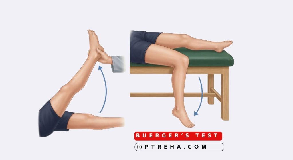
Buerger’s Test is a clinical bedside assessment used to evaluate arterial circulation in the lower limb. It is most commonly performed when peripheral arterial disease (PAD) or other arterial insufficiency is suspected, particularly when pulses are difficult to palpate or when the clinician wants a simple, low-cost indicator of limb perfusion.
What Is Buerger’s Test?
Buerger’s Test is a positional colour-change test that assesses the adequacy of arterial blood supply to the lower limb. It combines two observations: the development of pallor when the leg is elevated, and the development of reactive hyperaemia (rubor) when the leg is returned to a dependent position.
The test is named after Leo Buerger, who described it in the context of thromboangiitis obliterans, although the test itself assesses general arterial sufficiency rather than one specific disease. It is best classified as a clinical screening and observational assessment, not a provocative orthopedic special test and not a formal functional outcome measure.
Buerger’s Test provides a qualitative observation of changes in visible skin perfusion when the limb is elevated and then placed in a dependent position.. It does not measure ankle-brachial index, absolute perfusion pressure, tissue oxygenation, or the anatomical level of an arterial occlusion. It also does not distinguish between atherosclerotic PAD, thromboangiitis obliterans, embolic disease, or other causes of reduced arterial flow.
Its clinical role is to raise or lower suspicion of arterial insufficiency and to prompt further objective vascular testing when findings are abnormal.
Purpose of Buerger’s Test
- To screen for reduced arterial perfusion in the lower limb when arterial insufficiency is suspected.
- To support clinical reasoning when pedal pulses are diminished or difficult to palpate.
- To help differentiate, in combination with other findings, arterial from venous causes of lower limb symptoms.
- To provide a quick, equipment-free observation that can be repeated at the bedside or in clinic.
- To guide referral for more definitive vascular assessment such as ankle-brachial index, Doppler studies, or vascular imaging.
Buerger’s Test does not confirm a diagnosis on its own and should not be used as a substitute for objective vascular measurement.
Buerger’s Test Indications
- Patients reporting claudication, rest pain, cold feet, or non-healing wounds of the lower limb.
- Patients with risk factors for PAD, including diabetes, smoking history, hypertension, dyslipidaemia, or known cardiovascular disease.
- Patients in whom pedal pulses are absent or difficult to palpate.
- Patients with suspected thromboangiitis obliterans, particularly younger smokers with distal limb ischaemia.
- As part of a broader peripheral vascular examination in rehabilitation or primary care settings.
- When monitoring a patient with known arterial disease during a course of care, provided findings are interpreted cautiously.
The test is not appropriate for every patient with leg pain, and it should not be applied indiscriminately to patients with acute limb-threatening ischaemia.
Buerger’s Test Contraindications and Precautions
- Known or suspected acute critical limb ischaemia — do not delay urgent vascular referral to perform positional testing.
- Recent arterial or vascular surgery, or recent arterial catheterisation, unless cleared by the treating team.
- Unstable cardiac or respiratory status that limits safe leg elevation.
- Severe hip, knee, or ankle instability, recent lower limb fracture, or post-operative weight-bearing restrictions.
- Cognitive impairment or inability to follow positioning instructions, requiring modification or supervision.
- Patients with significant pain or who cannot tolerate limb elevation for the required observation period.
Modifications may include shorter elevation periods, supported positioning, or omission of the test in favour of non-positional vascular assessment when safety is uncertain.
Equipment
- A treatment plinth, examination couch, or bed that allows the leg to be elevated and then lowered.
- Pillows or wedges to support the limb in elevation if needed.
- A watch or timer for standardising observation periods.
- Adequate lighting to observe skin colour accurately.
- Gloves and a drape for patient comfort and dignity.
Patient Position
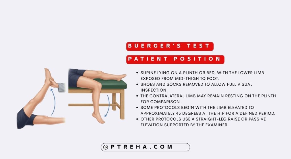
- Supine lying on a plinth or bed, with the lower limb exposed from mid-thigh to foot.
- Shoes and socks removed to allow full visual inspection.
- The contralateral limb may remain resting on the plinth for comparison.
- Some protocols begin with the limb elevated to approximately 45 degrees at the hip for a defined period.
- Other protocols use a straight-leg raise or passive elevation supported by the examiner.
Protocol variation exists between published descriptions, so the clinician should document the specific method used.
Examiner Position
- Standing at the side of the plinth, near the foot of the involved limb, with a clear view of the foot and toes.
- Positioned to support the limb during elevation if the patient cannot maintain the position independently.
- Guarding the limb to prevent sudden drops or uncontrolled movement during lowering.
- Avoiding pressure on the popliteal fossa or posterior thigh during elevation.
- Maintaining an unobstructed view of the plantar surface, dorsum, and toes for colour changes.
How to Perform Buerger’s Test
- Explain the procedure to the patient and obtain consent.
- Position the patient supine with both lower limbs exposed.
- Inspect and document baseline skin colour, temperature, capillary refill, and any wounds.
- Elevate the tested limb to approximately 45 degrees at the hip, or as tolerated, and hold for a standardised period (commonly around one to two minutes in published descriptions).
- Observe the foot and toes for the development of pallor. Note the timing and distribution of any colour change.
- Return the limb to a dependent position, either hanging over the edge of the plinth or lowered to the resting position.
- Observe for reactive hyperaemia — a dusky red or ruborous colour change — and note the time taken for colour to return.
- Record the presence, timing, and distribution of pallor and rubor, and compare with the contralateral limb where possible.
- Repeat only if clinically indicated, and document the protocol used.
There is no single universally accepted protocol for elevation angle, duration, or observation timing, so consistency within a clinic is important for meaningful comparison.
What Constitutes a Positive Test?
Some versions of the examination also record the vascular or Buerger angle—the degree of elevation at which pallor develops—but there is no universally validated angle that quantifies the severity or anatomical level of arterial obstruction.
Some versions of the examination also record the vascular or Buerger angle—the degree of elevation at which pallor develops—but there is no universally validated angle that quantifies the severity or anatomical level of arterial obstruction.”
The significance of these findings depends on the degree, timing, and distribution of colour change, and on the patient’s overall vascular presentation.
What Does an Abnormal Buerger’s Test Mean?
An abnormal finding suggests that arterial perfusion to the lower limb may be insufficient to meet positional demand. This is associated with conditions such as peripheral arterial disease and thromboangiitis obliterans, but it does not confirm either diagnosis.
Abnormal findings should prompt further assessment, including pulse palpation, ankle-brachial index, Doppler ultrasound, or vascular referral, depending on the clinical context.
Negative Buerger’s Test
A negative or normal Buerger’s Test means that no significant positional colour change was observed during the standardised protocol. It does not rule out arterial disease, particularly mild or well-compensated disease, and it does not exclude venous or neuropathic causes of symptoms.
A normal result should be interpreted alongside the full vascular examination and the patient’s history.
Reference Values and Cutoffs
There is no single universally accepted cutoff for Buerger’s Test. Findings are qualitative and depend on the elevation angle, duration, observation timing, lighting, skin pigmentation, and the examiner’s judgement.
Because of this variability, the test is best used as a supportive clinical observation rather than a graded measure with fixed thresholds.
Anatomy and Structures Involved in Buerger’s Test
- Arterial system: the femoral, popliteal, tibial, and pedal arteries supplying the lower limb.
- Skin and subcutaneous tissue: the visible vascular bed where colour change is observed.
- Microvascular circulation: reactive hyperaemia and changes in skin perfusion contribute to the dependent rubor observed after limb dependency.
- Hip and knee joints: involved in limb elevation and positioning.
Buerger’s Test is not a muscle-isolation test, and muscle contributions are relevant mainly to the positioning of the limb rather than to the assessment itself.
Conditions and Populations Associated With Buerger’s Test
- Peripheral arterial disease.
- Thromboangiitis obliterans (Buerger’s disease).
- Diabetes mellitus with peripheral vascular involvement.
- Chronic smokers with distal limb ischaemia.
- Patients with non-healing lower limb wounds or ulcers of suspected arterial origin.
- Patients being assessed for claudication or rest pain.
The test may raise or lower suspicion of these conditions but does not diagnose them.
Clinical Significance
- Provides a quick, low-cost observation of positional perfusion that can be performed in most clinical settings.
- Can support referral decisions when arterial insufficiency is suspected.
- Helps the clinician integrate subjective symptoms with objective bedside findings.
- May be repeated over time to monitor changes, though interpretation should remain cautious.
- Should always be combined with pulse palpation, capillary refill, ankle-brachial index, and other vascular assessments.
“Buerger’s Test is a bedside clue, not a verdict — it prompts further vascular assessment rather than replacing it.”
Reliability and Validity
Published evidence on the reliability and validity of Buerger’s Test is limited compared with objective vascular measures such as ankle-brachial index. Inter-rater reliability may be affected by differences in elevation angle, duration, lighting, and interpretation of colour change.
Validity is generally described in terms of association with arterial insufficiency rather than diagnostic accuracy. The test is therefore best regarded as a supportive clinical observation rather than a stand-alone measure.
Sensitivity and Specificity
Published estimates of sensitivity and specificity exist, but they are based on specific populations and protocols and should not be treated as universal diagnostic characteristics of the test.
Because of this variation, the test should not be presented as having fixed diagnostic accuracy, and it should not be used alone to confirm or exclude arterial disease.
Diagnostic Accuracy
Buerger’s Test is primarily a clinical screening and observational assessment. It should not be interpreted as a stand-alone diagnostic test. Diagnostic accuracy is better established for objective measures such as ankle-brachial index, Doppler studies, and vascular imaging.
Common Mistakes in Buerger’s Test
- Using inconsistent elevation angles or durations between assessments.
- Failing to standardise lighting, which affects colour interpretation.
- Not documenting baseline skin colour, temperature, or capillary refill.
- Interpreting colour change without considering skin pigmentation, ambient temperature, or venous status.
- Performing the test in patients with acute critical ischaemia, where urgent referral is the priority.
- Comparing findings across limbs without considering unilateral versus bilateral disease.
- Treating an abnormal finding as a confirmed diagnosis rather than a prompt for further assessment.
Tips for Performing Buerger’s Test
- Use consistent lighting and, where possible, the same room for repeat assessments.
- Standardise elevation angle and duration within your clinic and document them.
- Record baseline colour, temperature, and capillary refill before elevation.
- Observe the plantar surface and toes carefully, as changes may appear there first.
- Compare with the contralateral limb when both limbs are accessible.
- Document the timing and distribution of pallor and rubor, not just their presence.
- Combine findings with pulse palpation, ankle-brachial index, and history before drawing conclusions.
“Consistency in positioning and observation is what gives Buerger’s Test its clinical value; inconsistency turns it into guesswork.”
Limitations of Buerger’s Test
- It is qualitative and observer-dependent.
- It cannot identify the anatomical level or severity of arterial occlusion.
- Findings may be influenced by skin pigmentation, ambient temperature, venous disease, and oedema.
- Protocol variation limits comparability between clinicians and settings.
- It does not replace ankle-brachial index, Doppler studies, or vascular imaging.
- It should not be used in isolation to guide major clinical decisions.
Related Tests and Assessments
- Ankle-brachial index (ABI): provides an objective comparison of ankle and brachial systolic pressures and is an established non-invasive test for evaluating suspected PAD.
- Pulse palpation: assesses femoral, popliteal, dorsalis pedis, and posterior tibial pulses.
- Capillary refill time: provides a quick indicator of peripheral perfusion.
- Doppler ultrasound: evaluates arterial flow and can localise disease.
- Venous filling time: a separate peripheral vascular examination finding that may provide additional information about arterial insufficiency.
- Walking tests or treadmill testing: can assess functional walking limitation and, in appropriate patients, help evaluate exercise-related claudication
Frequently Asked Questions
Is Buerger’s Test a diagnostic test for peripheral arterial disease?
No. It is a clinical screening and observational assessment. It can raise suspicion of arterial insufficiency, but diagnosis requires objective vascular testing such as ankle-brachial index or imaging.
What is considered a normal Buerger’s Test result?
A normal result shows minimal or no pallor on elevation and prompt colour return on dependency. There is no universally accepted numerical threshold, and interpretation depends on the protocol and the patient’s baseline.
How long should the limb be elevated during Buerger’s Test?
Published protocols vary, but elevation is often maintained for around one to two minutes. The key is to use a consistent duration within your clinic and document it.
Can Buerger’s Test be used in patients with diabetes?
Yes, but findings should be interpreted cautiously. Diabetes can affect skin, microcirculation, and wound healing, and arterial disease may be more distal and diffuse. Objective vascular assessment remains important.
Does a negative Buerger’s Test rule out arterial disease?
No. Mild or well-compensated arterial disease may not produce obvious colour change. A negative result should be interpreted alongside the full vascular examination and history.
Is Buerger’s Test reliable between different examiners?
Inter-rater reliability is limited by variations in positioning, timing, lighting, and interpretation. Standardising the protocol within a clinic improves consistency.
When should Buerger’s Test be avoided?
It should be avoided or deferred in suspected acute critical limb ischaemia, recent vascular surgery, unstable cardiac or respiratory status, and situations where limb elevation is unsafe or contraindicated.
What should I do if Buerger’s Test is abnormal?
An abnormal finding should prompt further assessment, including pulse palpation, ankle-brachial index, Doppler studies, and appropriate vascular referral, depending on the clinical context.
Summary
- Buerger’s Test is a bedside observational assessment of lower limb arterial perfusion based on positional colour change.
- It involves elevating the limb to observe pallor, then lowering it to observe reactive hyperaemia.
- Findings are qualitative, and there is no universally accepted cutoff or scoring system.
- An abnormal finding suggests possible arterial insufficiency but does not confirm a diagnosis.
- Its principal limitation is that it cannot replace objective vascular measures such as ankle-brachial index or imaging.
Buerger’s Test remains a useful, low-cost component of the peripheral vascular examination when performed consistently and interpreted cautiously. Its greatest value lies in prompting further objective assessment rather than in providing a definitive answer on its own. Clinicians should document their protocol clearly, combine findings with other vascular tests, and refer appropriately when arterial insufficiency is suspected.