
The TMJ joint play test is a passive accessory-mobility assessment used within the physical examination of the temporomandibular joint (TMJ).
Rather than functioning as a stand-alone diagnostic test, it helps the clinician examine the quality and relative mobility of mandibular condylar movements, identify symptom reproduction, and determine whether restricted accessory motion may be relevant to the patient’s presentation.
Current physical-therapy literature considers accessory TMJ movements part of a broader examination that also includes mandibular range of motion, palpation, joint sounds, pain provocation, and cervical assessment.
What Is the TMJ Joint Play Test?
The TMJ joint play test is best classified as a clinical assessment of passive accessory joint mobility, rather than a conventional orthopedic special test with a universal positive or negative result.
Joint play refers to passive movement that occurs when an external force is applied to a joint.
For the TMJ, commonly described accessory movements include distraction, anterior or posterior translation, and medial or lateral gliding, with some clinical systems also using combined three-dimensional directions.
The examiner assesses characteristics such as relative excursion, resistance, end-feel, symmetry, movement quality, and whether the maneuver reproduces the patient’s familiar symptoms.
These observations are interpreted alongside the rest of the examination rather than in isolation.
Different manual-therapy approaches use different contact points, starting positions, and combinations of accessory directions.
Therefore, the exact technique should be documented rather than treating one procedure as the only universal version of a TMJ joint play examination.
Purpose of the TMJ Joint Play Test
The assessment may be used to:
- Examine the relative passive mobility of the mandibular condyle.
- Identify apparent restrictions in accessory translation or distraction.
- Compare mobility between the right and left TMJs when clinically appropriate.
- Observe resistance and end-feel during passive movement.
- Assess whether a particular accessory movement reproduces the patient’s familiar pain.
- Help determine whether an articular mobility impairment may be contributing to limited mandibular function.
- Guide clinical reasoning and selection of subsequent assessment or rehabilitation procedures.
The test is therefore more useful as part of a test cluster than as an isolated maneuver.
The DC/TMD literature specifically describes joint-play procedures as adjunctive tests rather than core diagnostic criteria.
TMJ Joint Play Test Indications
Joint play assessment may be considered when the patient presents with:
- Restricted or uncomfortable mouth opening.
- Suspected TMJ hypomobility.
- Pain apparently related to mandibular movement.
- A meaningful difference in mandibular movement between sides.
- Symptoms suggesting a possible intra-articular contribution.
- Persistent functional limitations involving chewing, yawning, speaking, or opening.
- A clinical question about whether restricted passive accessory motion is contributing to the patient’s movement impairment.
It can also be useful when the clinician needs to determine whether a previously identified movement limitation changes when accessory mobility is examined or addressed.
TMJ Joint Play Test Contraindications and Precautions
There is not a single universally established list of TMJ joint-play-specific contraindications.
Clinical judgment and the patient’s medical and dental history remain important.
Relevant precautions include:
- Obtain informed consent before external or intraoral manual examination.
- Use appropriate hand hygiene and gloves for intraoral techniques.
- Avoid unnecessarily forceful loading when the joint is acutely painful or highly irritable.
- Use particular care when the dentition is absent, sharp, damaged, or otherwise unsuitable for intraoral contact.
- Stop or modify the maneuver if it causes sharp, escalating, unfamiliar, or disproportionate symptoms.
- Consider gag sensitivity and patient apprehension when intraoral contact is required.
- Modify the examination when the patient cannot comfortably tolerate the proposed position or contact.
- Escalate or refer for further medical or dental evaluation when history or examination raises concern for pathology outside the scope of routine musculoskeletal assessment.
A useful principle is to distinguish symptom reproduction from simply creating discomfort through excessive manual force.
Patient Position
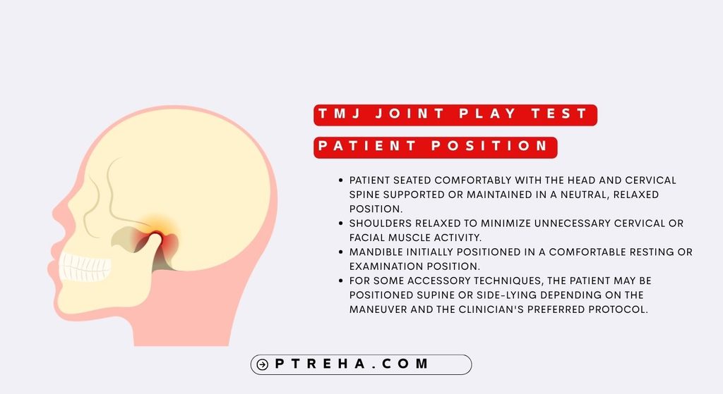
A commonly described position is:
- Patient seated comfortably with the head and cervical spine supported or maintained in a neutral, relaxed position.
- Shoulders relaxed to minimize unnecessary cervical or facial muscle activity.
- Mandible initially positioned in a comfortable resting or examination position.
- For some accessory techniques, the patient may be positioned supine or side-lying depending on the maneuver and the clinician’s preferred protocol.
The jaw may be examined at different degrees of opening because accessory mobility can vary with mandibular position.
Examiner Position
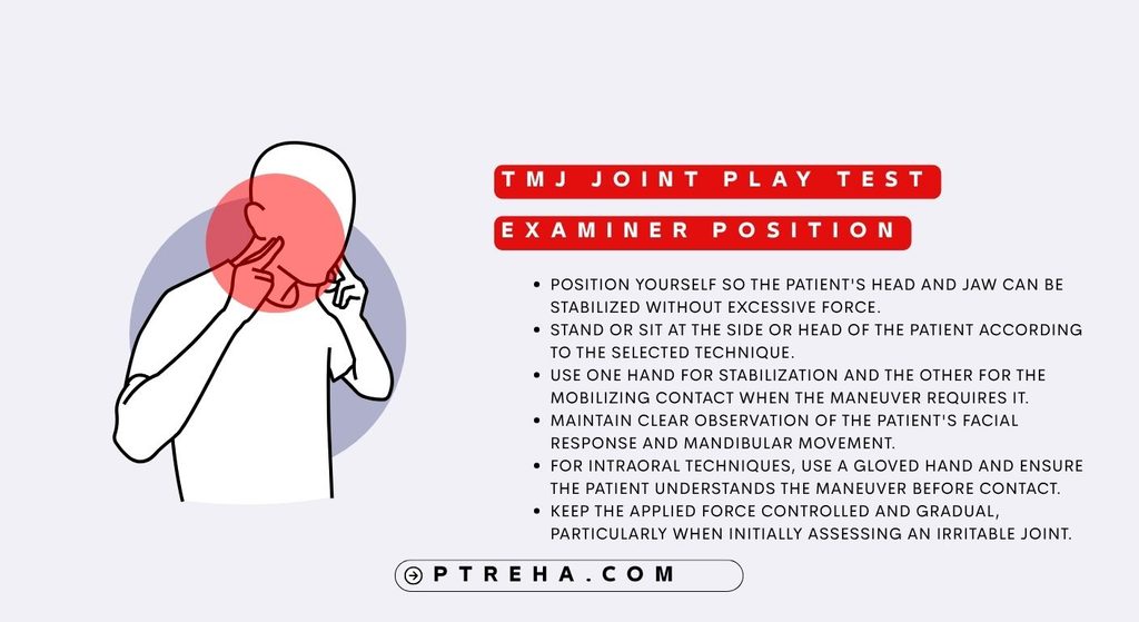
The examiner’s position depends on the specific accessory movement being tested.
- Position yourself so the patient’s head and jaw can be stabilized without excessive force.
- Stand or sit at the side or head of the patient according to the selected technique.
- Use one hand for stabilization and the other for the mobilizing contact when the maneuver requires it.
- Maintain clear observation of the patient’s facial response and mandibular movement.
- For intraoral techniques, use a gloved hand and ensure the patient understands the maneuver before contact.
- Keep the applied force controlled and gradual, particularly when initially assessing an irritable joint.
How to Perform the TMJ Joint Play Test
Because several accessory-mobility protocols are described in the literature, the following is a clinical examination framework, not a claim that there is only one standardized technique.
1. Establish the baseline
Begin with a history and observation of symptoms, then examine mandibular movements such as opening, protrusion, and lateral excursion.
Note whether movement is painful, restricted, asymmetric, or associated with joint sounds.
The DC/TMD examination emphasizes pain history, mandibular movements, joint sounds, and pain on palpation as part of the broader diagnostic examination.
2. Identify the TMJ and mandibular condyle
Palpate the TMJ region externally while the patient gently opens and closes the mouth.
Observe the timing and relative excursion of the mandibular condyles rather than assuming that a particular palpatory movement proves pathology.
Bilateral palpation can help the clinician compare the sides.
3. Assess distraction
For a commonly described distraction procedure:
- Stabilize the patient’s head.
- Place the gloved mobilizing hand around the mandible using an appropriate intraoral contact when the selected protocol calls for it.
- Apply a controlled caudal force to the mandible while maintaining an appropriate counterforce through the head or cranium.
- Palpate the joint region to appreciate the resulting relative separation or gapping motion.
- Record the quality of movement, resistance, symptom response, and side-to-side differences.
The purpose is to assess passive accessory mobility, not to force the joint through a large range.
4. Assess anterior glide
Anterior translation can be considered both dynamically and passively.
During active mandibular opening, protrusion, and lateral movement, the clinician can palpate the condyles externally to compare relative translation.
A passive version can then apply a gentle anterior-caudal force to the mandible while the joint is palpated.
The examiner should consider whether the condyle appears to translate relatively early, late, excessively, or insufficiently compared with the opposite side.
5. Assess medial and lateral glides
Medial and lateral accessory glides can be examined using external mandibular contacts with appropriate stabilization of the opposite side.
The examiner should assess:
- Relative movement quantity.
- Resistance.
- Movement quality.
- Symmetry.
- Familiar symptom reproduction.
Because the mandible is a coupled structure, stabilization of the opposite side is important when the clinician is attempting to emphasize movement on one side.
Complete mechanical isolation of one TMJ should not be assumed.
6. Consider combined three-dimensional movement
Some clinical approaches use a combined caudal-anterior-medial (CAM) direction to provide a three-dimensional assessment of accessory mobility.
This should be treated as a protocol-dependent examination strategy rather than a universally validated diagnostic maneuver.
7. Compare symptoms and movement
After the accessory examination, reassess the movement or functional activity that originally reproduced the patient’s symptoms.
A change in pain, ease of movement, or available motion can contribute to clinical reasoning, but a change after manual loading or mobilization does not by itself identify one specific anatomical lesion or establish a diagnosis.
Interpretation of the TMJ Joint Play Test
The TMJ joint play assessment does not have a universally accepted pass/fail criterion.
Clinicians commonly document:
- Restricted versus relatively unrestricted accessory motion.
- Symmetrical versus asymmetrical movement.
- Normal-feeling versus altered resistance or end-feel based on the examiner’s clinical assessment.
- Reproduction or non-reproduction of the patient’s familiar symptoms.
- The position in which restriction or symptoms occur.
- Whether the finding changes after another intervention or reassessment.
The most important distinction is between an observed impairment and a diagnostic conclusion.
For example, restricted passive accessory motion may support the hypothesis that joint mobility is contributing to a patient’s movement limitation, but it does not by itself establish osteoarthritis, disc displacement, synovitis, or another specific disorder.
Contemporary examination literature recommends integrating accessory findings with the remainder of the subjective and physical examination.
“Joint play is an assessment of mobility, not a diagnosis by itself.”
What Does Restricted TMJ Joint Play Mean?
A relatively restricted accessory movement may be clinically consistent with an articular mobility impairment, particularly when the patient also demonstrates restricted mandibular motion or pain during joint loading or movement.
However, several factors can influence the finding, including symptom irritability, guarding, examiner technique, mandibular position, bilateral coupling, and the patient’s ability to relax.
A restricted finding therefore warrants clinical correlation, not automatic labeling of a particular tissue lesion.
Conversely, relatively free accessory movement does not exclude a temporomandibular disorder.
The patient may have pain, disc-related symptoms, muscular pain, or other impairments despite apparently preserved accessory mobility.
Anatomy Involved in the TMJ Joint Play Test
Relevant structures include:
- Mandibular condyle: The articulating portion of the mandible assessed indirectly during accessory movement.
- Mandibular fossa and articular eminence: Components of the temporal bone forming the cranial articular environment of the TMJ.
- Articular disc: A fibrocartilaginous structure that contributes to the mechanics of mandibular movement.
- Joint capsule: Surrounds the joint and contributes to passive mechanical restraint.
- Temporomandibular ligament and associated capsular tissues: Contribute to passive stability and movement constraints.
- Retrodiscal tissues: Located posterior to the articular disc and relevant to symptom assessment in some patients.
- Masticatory musculature: Although not directly isolated by joint play, muscle guarding and activity can influence the clinical presentation and tolerance of passive movement.
- Cervical and craniofacial systems: These may contribute to the patient’s broader symptom presentation and should not be ignored during examination.
The manual procedure moves the mandible as a functional unit.
It should therefore not be described as a test that selectively isolates the capsule, disc, ligament, or an individual muscle.
Conditions Detected / Associated With the Test
The TMJ joint play examination may contribute information when evaluating presentations associated with:
- Temporomandibular disorders involving suspected articular impairment.
- Restricted mandibular mobility.
- TMJ-related pain or arthralgia as part of a broader examination.
- Joint hypomobility.
- Selected presentations involving altered accessory motion.
- Functional limitations where restricted TMJ mobility is part of the clinical impairment profile.
The test does not independently confirm a specific TMD diagnosis.
The DC/TMD framework uses a defined history and examination protocol for diagnosis, while joint-play procedures are considered adjunctive examinations.
Clinical Significance
The value of the TMJ joint play assessment is primarily its contribution to clinical reasoning.
- It can help determine whether passive accessory motion appears restricted.
- It may help identify whether symptoms are reproduced with particular directions of joint loading or movement.
- It can provide information that complements active mandibular range-of-motion testing.
- It may help the clinician decide whether further assessment of articular mobility is warranted.
- Reassessment after treatment can show whether a previously observed mobility impairment or functional limitation has changed.
- It can complement examination of the masticatory muscles and cervical spine rather than replacing them.
International physical-therapy consensus work identified accessory movements among the physical examination procedures considered clinically useful for patients with TMD.
Sensitivity and Specificity
A universal sensitivity or specificity value cannot be appropriately assigned to the TMJ joint play test.
The literature includes older diagnostic studies and more recent examination research, but the joint-play procedure is not a single standardized test with one universally accepted protocol and diagnostic cutoff.
The DC/TMD literature also identifies joint-play procedures as adjunctive rather than core diagnostic criteria.
For this reason, reporting one sensitivity or specificity number as though it applied to all patients and all joint-play techniques would be misleading.
Diagnostic Accuracy
Diagnostic accuracy is also protocol- and population-dependent and is not sufficiently established to treat the TMJ joint play test as an independent diagnostic test.
An older study found that the joint play test contributed some information when distinguishing arthrogenous subgroups, but active movement testing provided stronger discriminatory information, and the authors emphasized the value of the broader examination rather than reliance on one orthopedic test.
Contemporary diagnostic frameworks similarly place greater emphasis on a structured clinical examination rather than isolated accessory testing.
Reliability and Measurement Considerations
Recent research has specifically examined the reproducibility of TMJ joint-play assessment.
A study of 50 asymptomatic adults investigated several TMJ examination procedures, including joint play.
In that study, inter-rater agreement for joint mobility was relatively limited compared with some other examined procedures, indicating that examiner-dependent variation can matter.
The results should not be generalized to symptomatic patients or interpreted as a universal statement about every joint-play technique.
This is clinically important because joint play includes qualitative judgments about resistance, movement quality, and relative mobility.
Consistent positioning, contact, stabilization, force application, and documentation are therefore important when serial examinations are performed.
“Consistency of technique matters when a mobility finding is used to guide reassessment.”
Common Mistakes in the TMJ Joint Play Test
- Treating the procedure as a stand-alone diagnostic test.
- Calling a mobility finding universally “abnormal” without considering the patient’s presentation.
- Using excessive force to obtain a larger movement.
- Ignoring patient apprehension or guarding.
- Failing to compare symptoms with the patient’s familiar pain.
- Assuming that movement on one side can be completely isolated from the other side.
- Using a single accessory direction without considering other examination findings.
- Ignoring mandibular active range of motion, joint sounds, muscle palpation, or cervical findings.
- Using identical force or technique in every patient regardless of irritability.
- Recording only “restricted” without documenting direction, symptoms, resistance, and clinical context.
Tips for Performing the TMJ Joint Play Test
- Explain the maneuver before beginning, especially when intraoral contact is required.
- Start with low-force examination and increase only as clinically appropriate.
- Compare sides when comparison is meaningful.
- Assess the joint in more than one mandibular position when the clinical question requires it.
- Watch the patient’s facial expression and ask specifically about familiar versus unfamiliar symptoms.
- Keep stabilization controlled so that the movement being examined is not unnecessarily distorted.
- Document the exact accessory direction and position used.
- Reassess a relevant functional movement after the examination when appropriate.
- Use the finding to develop or test a clinical hypothesis rather than treating it as proof of a particular tissue lesion.
- Combine joint play findings with the patient’s history, active mandibular motion, palpation, symptom behavior, joint sounds, and relevant cervical examination.
Summary
The TMJ joint play test is a passive accessory-mobility clinical assessment used to examine the relative movement and mechanical behavior of the temporomandibular joint.
It commonly includes assessment of distraction, translation, and medial or lateral gliding, although exact techniques vary between clinical protocols.
Findings may include differences in mobility, resistance, movement quality, and reproduction of the patient’s familiar symptoms.
The examination can contribute to identifying a possible TMJ mobility impairment, but it does not independently diagnose a temporomandibular disorder or specify the exact anatomical lesion responsible for symptoms.
Interpretation should be integrated with active mandibular movement, pain provocation, palpation, joint-sound assessment, history, and other relevant findings.
Evidence regarding reproducibility and diagnostic usefulness is protocol-dependent, so clinicians should avoid universal cutoffs or isolated diagnostic conclusions.
A careful TMJ joint play assessment is most useful when it answers a specific clinical question: Is passive accessory mobility altered, does the movement reproduce the patient’s familiar symptoms, and does that information meaningfully change the clinical reasoning or subsequent management?
Resources and External Links
International consensus on physical-therapy assessment of TMD — von Piekartz H, Schwiddessen J, Reineke L, et al.
Journal of Oral Rehabilitation.
View the PubMed record
Evaluation and management of temporomandibular disorders: Part 1 — Shaffer SM, Naze G.
Journal of Manual & Manipulative Therapy.
View the PubMed record
Diagnostic Criteria for Temporomandibular Disorders (DC/TMD) — international diagnostic framework.
View the NIH resource
DC/TMD executive summary and diagnostic recommendations — diagnostic criteria and role of adjunctive joint-play testing.
Read the full article
Joint play movements of the temporomandibular joint: clinical considerations — Friedman MH, Weisberg J.
Archives of Physical Medicine and Rehabilitation.
View the PubMed record
Diagnostic value of orthopedic tests in patients with temporomandibular disorders — Lobbezoo-Scholte AM, Steenks MH, Faber JAJ, Bosman F.
Journal of Dental Research.
View the PubMed record
Reliability and minimal detectable change of common TMJ tests and measures — Olivencia O, Kaplan KB, Graham A, et al.
Physiotherapy Theory and Practice.
View the PubMed record