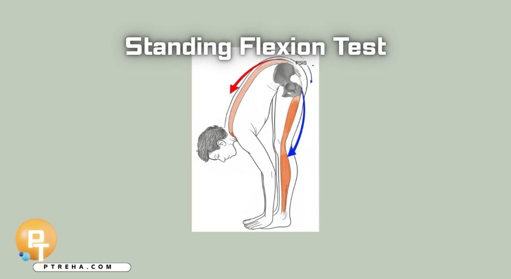
The Standing Flexion Test is a palpatory clinical assessment traditionally used in manual therapy to observe apparent asymmetry of pelvic landmark movement during forward trunk flexion. It is commonly associated with assessment of sacroiliac joint (SIJ) mobility, but its interpretation is limited because PSIS movement can be influenced by several factors and the test should not be used alone to diagnose SIJ dysfunction.
What Is the Standing Flexion Test?
The Standing Flexion Test, also called the standing forward flexion test or Vorlauf test, is a palpatory clinical assessment performed while the patient bends forward from standing.
Traditionally, the examiner palpates both posterior superior iliac spines (PSIS) and observes their relative movement during trunk flexion. Greater apparent cranial movement of one PSIS has traditionally been interpreted as an asymmetrical finding potentially related to SIJ motion.
However, this interpretation requires caution. The movement observed at the PSIS represents the combined result of trunk, lumbar spine, pelvis, hip, and soft-tissue movement rather than an isolated measurement of SIJ motion.
Recent literature continues to question whether the test can meaningfully identify SIJ dysfunction from palpatory movement alone.
Classification: The Standing Flexion Test is best considered a clinical palpatory assessment of pelvic/SIJ movement, rather than a standalone diagnostic test or outcome measure.
Purpose of the Standing Flexion Test
The assessment may be used to:
- Observe apparent asymmetry of PSIS movement during forward flexion.
- Complement a broader examination of the lumbopelvic region.
- Generate a clinical hypothesis about movement asymmetry.
- Compare findings with other examination procedures.
- Teach palpatory assessment and movement observation in manual therapy education.
- Contribute to clinical reasoning when interpreted alongside symptoms, functional findings, and other examination results.
It should not be treated as a standalone method for determining whether the SIJ is the source of a patient’s pain.
Standing Flexion Test Indications
The test may be considered when:
- A patient presents with lumbopelvic or posterior pelvic symptoms requiring physical examination.
- The clinician is assessing movement behavior of the pelvis during forward flexion.
- An examination includes palpatory assessment of the SIJ region.
- The clinician wants to compare the finding with other SIJ-related examination procedures.
- The assessment is being used for educational or movement-observation purposes.
The indication should come from the patient’s presentation and the clinical question rather than from the presence of an assumed SIJ dysfunction.
Standing Flexion Test Contraindications and Precautions
There are no universally established test-specific contraindications that should be presented as applying to every patient. Instead, the clinician should consider the patient’s general ability to safely perform forward trunk flexion.
Relevant precautions include:
- Significant acute pain that makes forward bending inappropriate.
- Recent trauma or surgery where unrestricted trunk or hip flexion has not been cleared.
- Conditions in which spinal or hip movement restrictions have been specifically prescribed.
- Dizziness, instability, or other symptoms that could make standing and bending unsafe.
- Neurological or musculoskeletal symptoms that worsen substantially with flexion.
The examination should be stopped if the maneuver produces concerning symptoms or the patient cannot perform it safely.
Patient Position
A commonly described protocol uses:
- The patient standing upright.
- Feet positioned approximately hip-width apart or in a comfortable standardized stance.
- Knees maintained in extension during the flexion movement.
- Arms positioned according to the chosen clinical protocol, commonly allowing the hands to move toward the floor.
- The patient instructed to bend forward slowly through the available range.
Different teaching and clinical protocols may vary, so the examiner should use one consistent procedure when comparing findings.
Examiner Position
A commonly described examiner position is:
- Stand behind the patient.
- Identify both PSIS by palpation.
- Place the thumbs or fingertips over the PSIS landmarks.
- Maintain light, consistent contact.
- Observe the relative movement of the landmarks as the patient flexes forward.
The examiner should avoid pressing excessively into the tissues because excessive palpatory pressure can alter the quality of the movement being assessed.
How to Perform the Standing Flexion Test
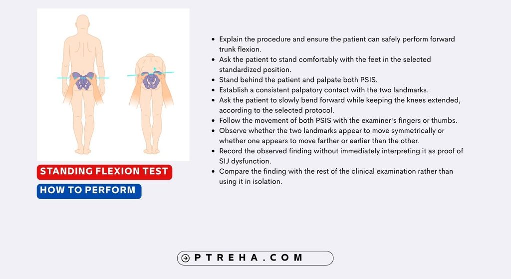
- Explain the procedure and ensure the patient can safely perform forward trunk flexion.
- Ask the patient to stand comfortably with the feet in the selected standardized position.
- Stand behind the patient and palpate both PSIS.
- Establish a consistent palpatory contact with the two landmarks.
- Ask the patient to slowly bend forward while keeping the knees extended, according to the selected protocol.
- Follow the movement of both PSIS with the examiner’s fingers or thumbs.
- Observe whether the two landmarks appear to move symmetrically or whether one appears to move farther or earlier than the other.
- Record the observed finding without immediately interpreting it as proof of SIJ dysfunction.
- Compare the finding with the rest of the clinical examination rather than using it in isolation.
A key issue is consistency. Differences in foot position, knee flexion, speed of movement, palpation pressure, trunk strategy, or examiner technique can alter the observed result.
Test Findings and Interpretation
Unlike a conventional outcome measure with a numerical score, the Standing Flexion Test is generally interpreted through qualitative observation of relative PSIS movement.
A commonly described finding is that one PSIS appears to move farther cranially than the other during forward flexion. Traditional manual-therapy approaches may describe this as an asymmetrical or positive finding.
However, the clinically important point is that asymmetrical PSIS movement does not establish SIJ dysfunction.
The observed movement can be affected by multiple components of the movement system, including lumbar and hip motion, pelvic morphology, muscle tension, and the mechanics of the patient’s forward-flexion strategy.
“An asymmetrical palpatory finding is a clinical observation, not a diagnosis.”
For this reason, it is more appropriate to document what was observed rather than automatically labeling one side as “dysfunctional.”
What Does an Asymmetrical Finding Mean?
An asymmetrical finding means that the examiner perceived a difference in the movement of the two PSIS landmarks during the selected flexion maneuver.
It does not independently establish:
- SIJ pain as the source of symptoms.
- SIJ hypomobility.
- A specific pelvic positional fault.
- A specific anatomical lesion.
- The need for a particular manual therapy technique.
The finding should instead be integrated with the patient’s history, symptom behavior, active movement examination, neurological examination when indicated, hip assessment, and other relevant clinical tests.
Anatomy Involved in Standing Flexion Test
Relevant structures and systems include:
- Posterior superior iliac spines (PSIS): palpable pelvic landmarks used as reference points during the assessment.
- Ilium: contributes to the movement of the pelvic landmarks being palpated.
- Sacroiliac joints: traditionally considered the primary joints being assessed.
- Sacrum: forms the other component of the SIJ.
- Lumbar spine: trunk flexion involves lumbar movement and therefore influences pelvic movement.
- Hip joints: hip flexion contributes substantially to the overall forward-bending movement.
- Hamstrings and other posterior-chain tissues: their length and tension can influence the patient’s flexion strategy.
- Trunk and pelvic musculature: coordinated activity contributes to the movement pattern.
The test therefore reflects a multijoint movement, rather than isolating SIJ movement.
Conditions Detected / Associated With the Test
The Standing Flexion Test has traditionally been associated with:
- Sacroiliac joint movement assessment.
- Suspected SIJ dysfunction.
- Lumbopelvic movement asymmetry.
However, the evidence does not support treating the test as an independent diagnostic test for these conditions. A systematic review found substantial uncertainty surrounding the validity of palpatory SIJ mobility tests, and a recent critical review highlights continuing controversy regarding the clinical meaning of Standing Flexion Test findings.
This distinction is particularly important when documenting clinical findings: “asymmetrical PSIS movement during standing flexion” is more defensible than automatically documenting “SIJ dysfunction.”
Clinical Significance
The Standing Flexion Test can still have a role within a broader physical examination.
Its potential clinical value includes:
- Providing an additional observation of lumbopelvic movement.
- Identifying movement asymmetry that may warrant further examination.
- Encouraging comparison between observed movement and the patient’s symptoms.
- Supporting clinical reasoning when combined with other findings.
- Providing a reproducible palpatory procedure for educational purposes.
Its limitations are equally important. A 2021 systematic review of palpatory SIJ mobility tests found that concurrent validity had not been established in the included studies and concluded that further research was needed.
Reliability and Validity Evidence
Reliability findings for the Standing Flexion Test are protocol- and study-dependent.
An older study involving asymptomatic participants reported poor inter-examiner agreement but higher intra-examiner agreement, illustrating how examiner-related variability can influence the result.
A 2021 systematic review and meta-analysis reported moderate intra-examiner agreement for the Standing Flexion Test, but concluded that further research was necessary because concurrent validity had not been established for the palpatory SIJ mobility tests examined.
Another 2021 study examining the Standing Flexion Test in healthy participants reported construct-validity and intra-rater findings but very low inter-rater agreement. Importantly, its sample consisted of healthy individuals, so the results should not automatically be generalized to patients with low back or SIJ-related symptoms.
More recent research continues to investigate whether standardized training can improve reproducibility, reinforcing that examiner technique and protocol standardization remain important issues.
Sensitivity and Specificity
Sensitivity and specificity estimates reported in the literature are not sufficiently consistent to provide a universal value for the Standing Flexion Test.
Different studies use different populations, reference standards, procedures, and definitions of an abnormal finding. Therefore, a single sensitivity or specificity value should not be presented as representative of the test in general.
Diagnostic Accuracy
The Standing Flexion Test should not be considered a standalone diagnostic test for SIJ dysfunction.
The principal limitation is that apparent PSIS movement is influenced by the movement of multiple anatomical regions and by palpatory judgment. Contemporary reviews therefore continue to question whether palpatory movement tests can accurately identify SIJ dysfunction on their own.
A useful clinical distinction is:
The test can provide an examination finding; the finding does not by itself provide a diagnosis.
Common Mistakes in Standing Flexion Test
- Treating an asymmetrical PSIS movement as definitive evidence of SIJ dysfunction.
- Using inconsistent foot positions between examinations.
- Allowing the knees to flex during one assessment but not another.
- Applying excessive pressure with the thumbs.
- Losing contact with the PSIS during the movement.
- Moving the patient’s pelvis manually while attempting to palpate it.
- Performing the test too quickly.
- Ignoring hip or lumbar contributions to the movement.
- Assuming that a visible or palpable asymmetry identifies the painful structure.
- Using the result without considering the patient’s symptoms and complete examination.
Tips for Performing the Standing Flexion Test
- Explain the movement clearly before starting.
- Use the same procedure when reassessing the patient.
- Establish both PSIS landmarks before asking for flexion.
- Maintain light and consistent palpatory contact.
- Ask for slow, controlled forward flexion.
- Observe the entire movement rather than focusing exclusively on the end position.
- Consider lumbar and hip movement when interpreting the observation.
- Record the actual finding rather than immediately assigning a pathological label.
- Combine the result with other clinically relevant examination findings.
- Avoid using the test as the sole basis for treatment selection.
“Use the Standing Flexion Test to add information to an examination, not to replace the examination.”
Summary
- The Standing Flexion Test is a palpatory clinical assessment traditionally used to observe relative PSIS movement during forward trunk flexion.
- It is commonly associated with assessment of sacroiliac joint mobility, but it does not independently establish SIJ dysfunction or identify the source of pain.
- The observed movement is influenced by the lumbar spine, hips, pelvis, soft tissues, movement strategy, and examiner technique.
- Research has reported variable reliability, with particularly important concerns regarding inter-examiner agreement and clinical validity.
- The result is most appropriately interpreted as one finding within a comprehensive physical therapy assessment.
The Standing Flexion Test can be useful as a structured observation of lumbopelvic movement, particularly in education and clinical examination. Its greatest limitation is also its most important teaching point: an asymmetrical palpatory finding should prompt further clinical reasoning, not an automatic diagnosis of SIJ dysfunction.
Resources and External References
- PubMed — Validity and Reliability of Palpatory Clinical Tests of Sacroiliac Joint Mobility: A Systematic Review and Meta-analysis
- PubMed — Construct Validity and Reliability of Tests for Sacroiliac Dysfunction: Standing Flexion Test and Sitting Flexion Test
- PubMed — Inter-examiner and Intra-examiner Reliability of the Standing Flexion Test
- PMC — The Standing Forward Flexion Test in Manual Therapy: A Critical Review and a Functional Reinterpretation
- PMC — Improving the Reproducibility of the Standing Forward Flexion Test After Specific Training
- Journal of Osteopathic Medicine — Construct Validity and Reliability of Tests for Sacroiliac Dysfunction