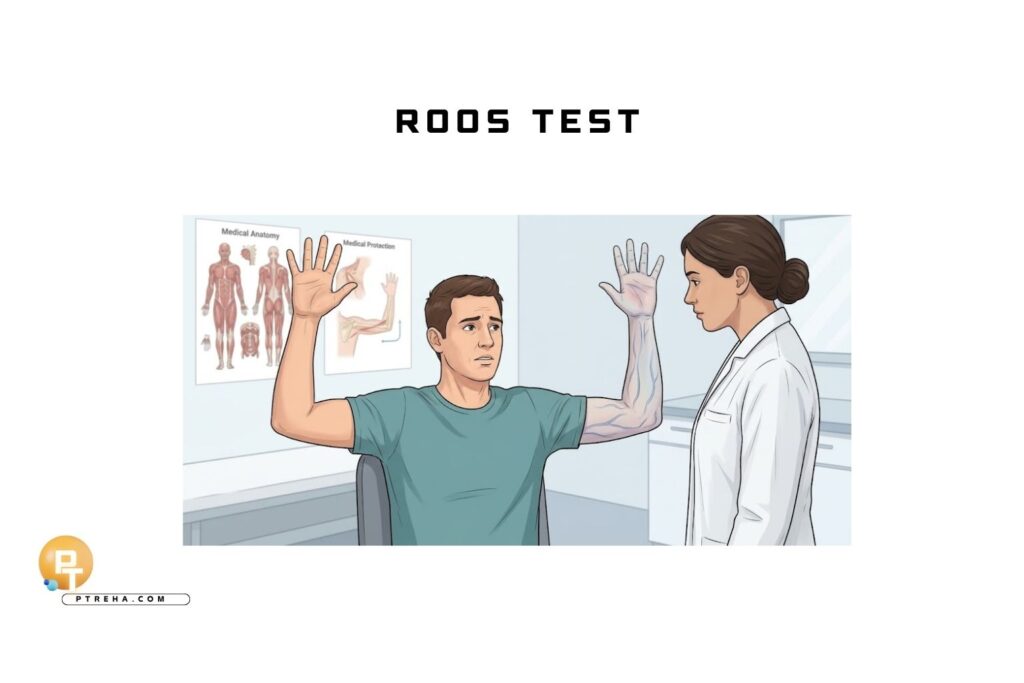
The Roos Test assesses whether sustained arm elevation and repeated hand movements reproduce a patient’s upper-limb symptoms.
Also called the elevated arm stress test (EAST), it can contribute to assessment for thoracic outlet syndrome, but its findings require careful interpretation alongside the history and wider examination.
What Is the Roos Test?
The Roos Test is a provocative orthopedic special test, commonly used in the clinical assessment of suspected neurogenic thoracic outlet syndrome (NTOS).
It evaluates symptom reproduction during a sustained position; it does not directly measure nerve compression, blood flow, or isolated muscle strength.
The INTOS consensus recommendations for neurogenic thoracic outlet syndrome recommend EAST as a provocative maneuver within a broader diagnostic assessment.
This recommendation does not establish it as an independently diagnostic test.
Purpose of the Roos Test
- Explore whether arm elevation reproduces the patient’s familiar pain, tingling, numbness, or perceived weakness.
- Compare the symptoms elicited during examination with those reported during overhead activities.
- Contribute to clinical reasoning when NTOS is suspected.
Before testing, establish what the patient means by “weakness” or “numbness.” A familiar hand symptom is clinically different from ordinary shoulder fatigue during sustained elevation.
Roos Test Indications
Consider the test when the history suggests:
- Upper-limb symptoms aggravated by elevated arm positions.
- Pain, paresthesia, or perceived weakness during repetitive arm or hand use.
- Possible NTOS requiring assessment alongside alternative explanations.
The test should answer a question arising from the history.
It should not be used as an isolated screening tool to label an otherwise asymptomatic person with thoracic outlet syndrome.
Roos Test Contraindications and Precautions
A universally accepted list of Roos-specific contraindications is not established.
Apply the usual precautions for a sustained, symptom-provoking upper-limb examination.
- Defer testing and arrange urgent assessment when there is suspected acute vascular compromise, such as sudden arm swelling or a cold, pale, painful hand.
- Respect movement restrictions following injury or surgery; do not force the test position.
- If shoulder pain or restricted movement prevents positioning, document the limitation rather than treating an incomplete test as evidence of NTOS.
- Stop for intolerable pain, rapidly worsening neurological symptoms, marked color change, or the patient’s request.
These are clinical safety decisions, not validated Roos-specific exclusion criteria.
Do not continue simply to complete the planned test duration.
Patient Position
For the commonly described three-minute EAST protocol:
- Position both shoulders in approximately 90° of abduction and external rotation.
- Flex both elbows to approximately 90°, with the upper arms at shoulder height.
- Keep the arms unsupported during the maneuver.
These angles describe the protocol, not diagnostic thresholds.
The clinical overview of NTOS assessment describes this position with repeated hand opening and closing for three minutes.
Positioning and execution vary in practice; record the version used.
Examiner Position
- Stand or sit where both arms, hands, and the patient’s expression are visible.
- Check the starting position and observe any loss of position.
- Keep a timer available and remain close enough to end the maneuver promptly.
- Avoid supporting the arms or imposing additional shoulder depression.
How to Perform the Roos Test
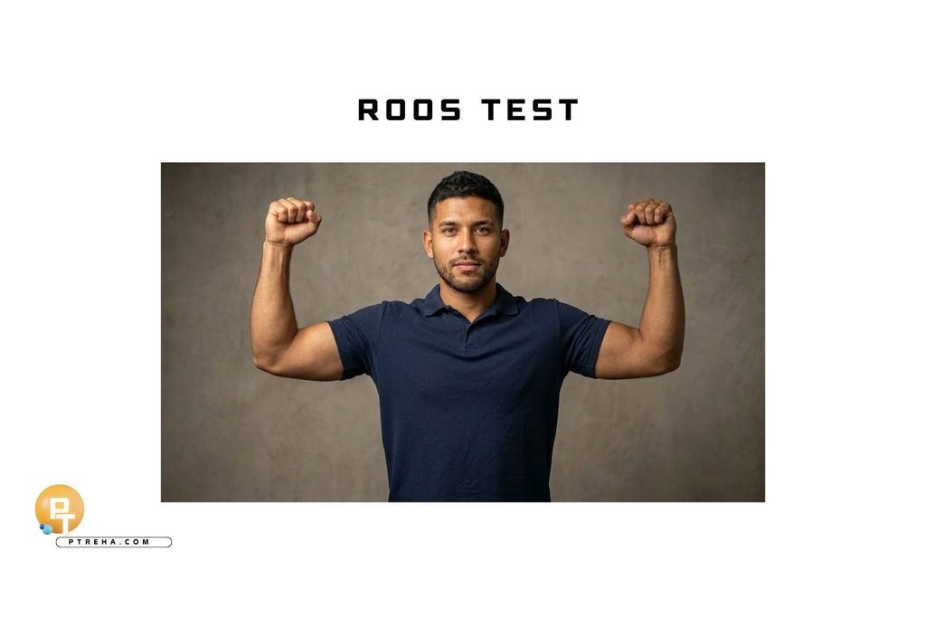
- Record baseline symptoms. Ask about their location, character, and severity, including whether they are already present at rest.
- Explain the maneuver. Ask the patient to report symptom onset and whether the sensation matches their usual complaint.
- Demonstrate the arm position. Confirm that the patient can assume it without being forced.
- Begin repeated hand movements. In the protocol described above, the patient opens and closes both hands at approximately one cycle per second.
- Continue for up to three minutes, ending earlier if symptoms or safety concerns require stopping.
- Observe and document. Record familiar symptom reproduction, laterality, onset time, changes in arm position, and the reason for stopping.
- Reassess after lowering the arms. Record whether symptoms settle and whether any neurological or vascular concern remains.
The three-minute duration is the planned endpoint of this protocol.
It is not a universal pass/fail threshold.
What Constitutes a Positive Test?
The Roos Test is commonly described as positive when it reproduces symptoms compatible with the patient’s suspected thoracic outlet presentation.
For suspected NTOS, recognizable pain, paresthesia, or perceived weakness is more informative than a vague report of tired arms.
Criteria vary between descriptions.
Inability to continue may accompany symptom provocation, but stopping early because of shoulder discomfort, general fatigue, or difficulty following instructions should not automatically be labeled positive for NTOS.
Document the actual response rather than writing only “Roos positive.”
“Record the familiar symptom that was reproduced, not just whether the arms became tired.”
What Does a Positive Roos Test Mean?
A positive response may support suspicion of NTOS when it fits the history and other examination findings.
It does not establish the diagnosis, locate a specific compression site, or distinguish neural symptoms from vascular disease by itself.
For example, reproduction of the patient’s usual hand tingling during elevation warrants further assessment.
It does not identify which nerve structure is affected or prove that compression occurred at the thoracic outlet.
Alternative explanations remain relevant, including cervical radiculopathy, distal nerve entrapment, and shoulder disorders.
More than one condition can coexist.
Negative Roos Test
A negative result generally means that the chosen protocol did not reproduce the relevant symptoms.
It does not reliably exclude NTOS.
Record whether the patient completed the intended position and duration.
A modified or prematurely terminated test provides different information from a fully completed maneuver without symptom reproduction.
Anatomy Involved in Roos Test
Relevant anatomy includes:
- Brachial plexus: the neural structure involved in neurogenic thoracic outlet presentations.
- Subclavian artery and vein: relevant to arterial and venous thoracic outlet conditions.
- Scalene triangle, costoclavicular space, and retropectoralis minor region: anatomical regions considered during thoracic outlet assessment.
- Shoulder girdle and hand musculature: active during sustained elevation and repeated hand movement, contributing to the effort required.
The maneuver does not isolate one of these structures.
Symptoms during testing cannot, by themselves, identify a compression site.
Conditions Associated With the Test
- Suspected NTOS: the main clinical context for the symptom-provoking EAST described here.
- Possible vascular thoracic outlet presentations: vascular symptoms require appropriate medical evaluation; EAST does not confirm arterial or venous disease.
- Other upper-limb disorders: cervical, shoulder, or peripheral nerve conditions may explain symptoms elicited during testing.
A radial pulse change alone should not be used to diagnose NTOS.
Clinical Significance
- Interpret familiar symptom reproduction alongside the history and neurological, cervical, and shoulder examination.
- Use the response to guide further assessment rather than to select treatment automatically.
- Treat objective weakness, muscle wasting, or concerning vascular findings as reasons for further medical assessment.
- If repeating the maneuver, use the same setup and document changes in symptoms and execution.
Improved tolerance on a later examination does not, by itself, demonstrate that nerve compression has resolved.
Pair observations with meaningful changes in daily activity and other examination findings.
Sensitivity, Specificity, and Diagnostic Accuracy
There is no single sensitivity or specificity estimate that can be applied universally across Roos Test protocols and thoracic outlet populations.
Interpretation depends on how a positive response is defined and how the diagnosis is established.
In research examining EAST duration in patients referred with suspected NTOS, duration showed limited ability to distinguish those classified as having NTOS from symptomatic controls.
This finding concerns duration-based assessment in that clinical cohort; it should not be converted into a universal stopping-time cutoff.
Reliability and diagnostic accuracy are different properties.
A repeatable finding can still have limited ability to identify a particular condition.
Common Mistakes in Roos Test
- Treating nonspecific fatigue as confirmation of NTOS.
- Failing to ask whether the provoked symptoms are familiar.
- Allowing the arms to drift without documenting the change.
- Adding forced positioning or resistance to the described protocol.
- Recording only “positive” or “negative,” without the actual findings.
- Assuming that a negative result excludes NTOS.
Tips for Performing the Roos Test
- Demonstrate the movement before starting the timer.
- Use neutral questions, such as “What do you feel, and where?”
- Ask separately about shoulder effort and distal neurological symptoms.
- Record why the test ended, especially when symptoms were not the reason.
- Allow provoked symptoms to settle before another maneuver, where clinically appropriate.
Summary
- The Roos Test is a provocative special test used within assessment for suspected NTOS.
- A commonly described protocol uses elevated, externally rotated shoulders, flexed elbows, and repeated hand movements for up to three minutes.
- Familiar symptom reproduction is more informative than nonspecific fatigue.
- Positive and negative findings must be interpreted alongside the history and wider examination.
- The test cannot independently confirm or exclude NTOS or identify a compression site.
Use the Roos Test to explore the patient’s symptom behavior, document the response precisely, and decide what needs further assessment.
Its value comes from thoughtful clinical interpretation rather than a stand-alone label.