The McMurray Test is an orthopedic special test used during knee examination when a clinician suspects meniscal involvement.
In physical therapy practice, it is most useful as one part of a broader assessment that includes the injury mechanism, symptoms, joint-line examination, range of motion, functional findings, and other appropriate tests rather than as a stand-alone method for diagnosing a meniscal tear.
What Is the McMurray Test?
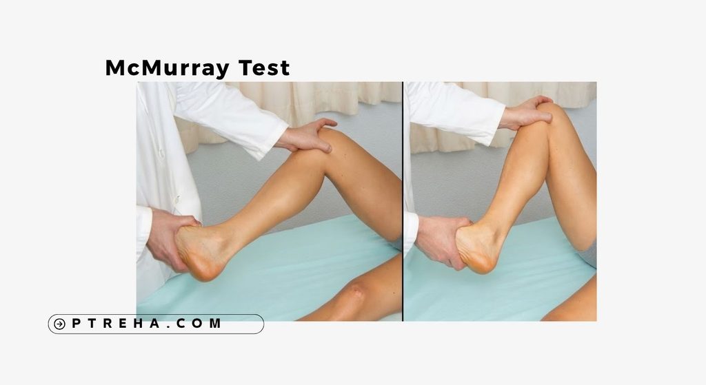
The McMurray Test is a provocative orthopedic special test used to assess whether a patient’s knee symptoms and mechanical findings are consistent with meniscal pathology.
The examiner moves the knee through flexion and extension while combining tibial rotation with a varus or valgus-directed force.
This maneuver changes loading and movement at the tibiofemoral joint and may reproduce symptoms or a palpable mechanical finding in a knee with meniscal pathology.
The test does not directly visualize the meniscus, determine the precise tear morphology, or establish a diagnosis by itself.
In clinical practice, I treat the McMurray Test as one piece of the examination rather than a final answer.
A test finding becomes much more meaningful when it agrees with the patient’s history, location of symptoms, mechanical complaints, and other examination findings.
The American Academy of Orthopaedic Surgeons’ guidance on acute isolated meniscal pathology includes the McMurray Test, joint-line tenderness, and Thessaly Test as components of physical examination and notes that examination findings may be more useful when considered together.
“A McMurray finding should change your clinical reasoning, not replace it.”
Purpose of the McMurray Test
The primary purpose is to provoke findings that may be associated with a meniscal injury during a structured knee examination.
The McMurray Test may help the clinician:
- Assess a patient with suspected meniscal involvement.
- Attempt to reproduce familiar joint-line symptoms.
- Identify a palpable or audible mechanical event associated with the maneuver.
- Compare examination findings with the patient’s history.
- Examine suspected medial or lateral meniscal involvement.
- Add information to other knee examination findings.
- Determine whether further evaluation may be appropriate.
- Build a clinical hypothesis rather than establish a diagnosis in isolation.
It should therefore be regarded as an adjunct to clinical assessment, not as a substitute for a complete knee evaluation.
McMurray Test Indications
The test may be considered when the history and examination reasonably suggest meniscal involvement.
Examples include:
- Knee pain following a twisting or rotational injury.
- Pain localized around the medial or lateral joint line.
- Mechanical catching or clicking associated with knee movement.
- A history of intermittent knee locking or restricted movement.
- Symptoms associated with loaded rotation or deep knee flexion.
- Suspicion of an acute traumatic meniscal injury.
- Persistent joint-line symptoms requiring further clinical assessment.
However, symptoms such as clicking or joint-line pain are not specific to meniscal tears.
Similar complaints can occur with other intra-articular or periarticular knee conditions.
The patient’s overall presentation should therefore determine whether the McMurray Test is appropriate.
McMurray Test Contraindications and Precautions
There is no universally accepted list of McMurray-specific absolute contraindications that applies to every patient.
The important issue is whether performing a provocative knee maneuver is clinically safe and necessary.
Relevant precautions include:
- Severe acute pain that prevents safe examination.
- Marked swelling or irritability that substantially limits movement.
- A knee that is mechanically locked and cannot safely be moved through the required range.
- Suspected fracture or significant acute structural injury requiring further medical assessment.
- Recent major trauma in which provocative testing is inappropriate.
- Inability of the patient to relax sufficiently for controlled passive testing.
- Symptoms that worsen substantially during positioning.
- Postoperative restrictions that prohibit the required movement or loading.
- Any situation in which the examination maneuver would add unnecessary risk without useful clinical information.
Do not force the knee through painful or mechanically blocked movement merely to obtain a test result.
When the patient’s knee is highly irritable, history, observation, range-of-motion assessment, and less provocative examination procedures may provide more useful information initially.
Patient Position
A commonly described McMurray Test protocol uses the following starting position:
- The patient lies supine.
- The hip is flexed sufficiently to allow the examiner to control the leg comfortably.
- The knee is brought into substantial flexion.
- The patient is encouraged to relax the lower limb.
- The heel or distal leg is supported by the examiner.
- The examiner’s other hand monitors the knee and joint line.
Exact hand placement and the degree of starting flexion vary somewhat among descriptions of the test.
This variability is clinically important because differences in technique and interpretation can affect test performance.
Examiner Position
The examiner generally stands beside the involved leg.
A practical setup includes:
- One hand controlling the heel or distal lower leg.
- The other hand positioned around the knee.
- Fingers placed near the joint line when possible.
- The examiner maintaining control of tibial rotation.
- The forearm or hand position allowing controlled varus or valgus stress.
- The leg remaining supported throughout the maneuver.
The movement should be deliberate and smooth rather than forceful.
A rushed McMurray Test often becomes difficult to interpret because the clinician may be unable to determine whether the patient’s response came from tibiofemoral rotation, excessive pressure, patellofemoral movement, or another source.
How to Perform the McMurray Test
Several descriptions and modified versions of the McMurray Test exist.
The following represents a commonly used clinical approach, not the only valid protocol.
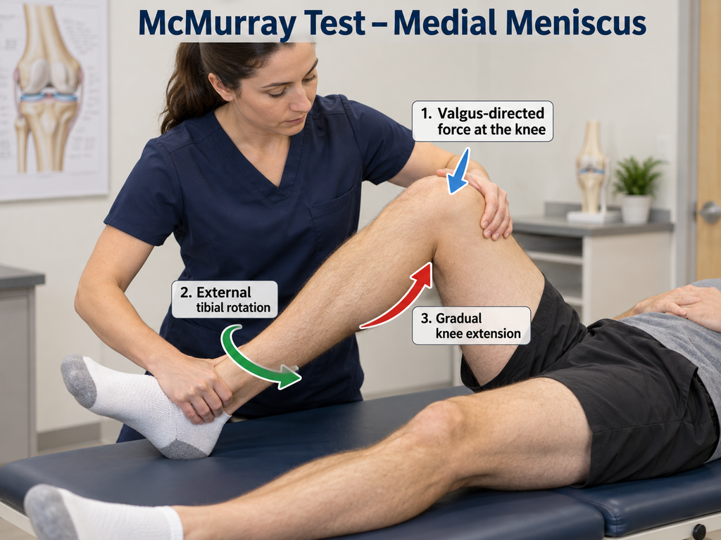
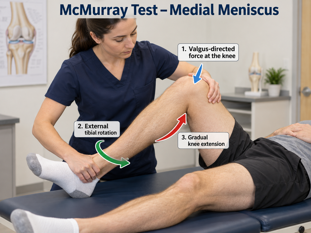
Assessing the Medial Meniscus
- Position the patient supine.
- Flex the hip and knee while supporting the leg.
- Bring the knee into substantial flexion as tolerated.
- Externally rotate the tibia relative to the femur.
- Apply a controlled valgus-directed force at the knee.
- Maintain the rotation while gradually extending the knee.
- Palpate the medial joint line during the movement.
- Ask whether the maneuver reproduces the patient’s familiar symptoms.
- Observe and feel for a mechanical click or clunk.
- Stop if the maneuver produces excessive pain or feels unsafe.
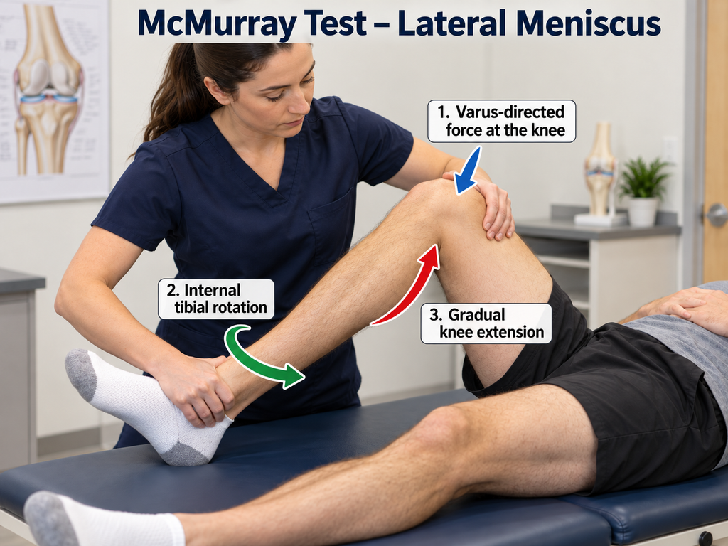
Assessing the Lateral Meniscus
The maneuver is generally reversed:
- Begin with the knee flexed.
- Internally rotate the tibia relative to the femur.
- Apply a controlled varus-directed force.
- Gradually extend the knee while maintaining controlled rotation.
- Monitor the lateral joint line.
- Note reproduction of recognizable symptoms or a mechanical finding.
Clinicians sometimes repeat the movement through different portions of knee flexion because different parts of the meniscus may be stressed as tibiofemoral contact changes.
Repeated aggressive cycling of a painful knee is unnecessary.
What Constitutes a Positive McMurray Test?
The terms positive and negative are commonly used for the McMurray Test, so they are clinically appropriate provided their limitations are understood.
Traditionally, a palpable or audible click, clunk, or similar mechanical event occurring during the maneuver has been considered a positive finding, particularly when it corresponds to the symptomatic joint line.
Some clinical protocols and research studies also incorporate reproduction of joint-line pain into their definition of a positive test.
This creates an important problem: not every study defines a positive McMurray Test in exactly the same way.
A useful examination record should therefore state what actually occurred rather than documenting only “McMurray positive.”
For example:
- Medial joint-line pain reproduced during external tibial rotation and extension.
- Palpable medial joint-line click with familiar pain.
- Painless click without reproduction of the patient’s symptoms.
- Lateral joint-line pain without a palpable mechanical event.
This documentation gives another clinician considerably more information than the word “positive.”
What Does a Positive McMurray Test Mean?
A positive McMurray Test can increase suspicion of meniscal pathology, particularly when the finding is consistent with the mechanism of injury, joint-line symptoms, and other examination findings.
It does not establish:
- The presence of a tear with certainty.
- The exact location of a tear.
- Tear size.
- Tear morphology.
- Whether a tear is traumatic or degenerative.
- Whether the meniscal finding is actually responsible for all the patient’s symptoms.
- Whether surgical treatment is required.
This distinction is particularly important in adults who may have meniscal abnormalities on imaging without those abnormalities being the primary source of symptoms.
The McMurray Test therefore contributes to a clinical probability, not a stand-alone tissue diagnosis.
Negative McMurray Test
A negative McMurray Test means that the selected maneuver did not reproduce the predefined finding used by that examiner or protocol.
A negative result does not reliably exclude a meniscal tear.
This is one of the major limitations of the test.
If the history strongly suggests meniscal involvement but McMurray testing is negative, the clinician should not automatically abandon that hypothesis.
Instead, consider:
- Mechanism of injury.
- Joint-line tenderness.
- Effusion.
- Range-of-motion limitations.
- Mechanical symptoms.
- Other meniscal provocative tests.
- Ligament examination when appropriate.
- Functional examination.
- The need for additional medical evaluation or imaging.
The systematic-review literature has repeatedly shown substantial variation in the sensitivity of McMurray testing, reinforcing why a negative result cannot safely be treated as definitive.
“A negative McMurray Test does not erase a convincing clinical history.”
Anatomy Involved in the McMurray Test
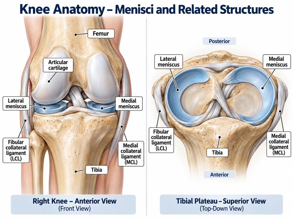
Understanding the anatomy helps explain why tibial rotation, knee flexion, and extension are incorporated into the maneuver.
Relevant structures include:
- Medial meniscus: fibrocartilaginous structure between the medial femoral condyle and medial tibial plateau.
- Lateral meniscus: fibrocartilaginous structure between the lateral femoral condyle and lateral tibial plateau.
- Femoral condyles: articulate with the tibia and menisci during knee movement.
- Tibial plateau: supports the menisci and participates in tibiofemoral articulation.
- Joint capsule: surrounds the knee joint and contributes to joint stability.
- Meniscocapsular attachments: connect portions of the menisci to surrounding capsular structures.
- Collateral ligament structures: relevant to the forces introduced during valgus- and varus-directed components of the maneuver.
- Tibiofemoral joint: the articulation in which the rotational and flexion-extension components occur.
The menisci contribute to load distribution, joint congruence, shock attenuation, and knee mechanics.
During the McMurray maneuver, changing knee flexion and tibial rotation changes the mechanical relationship between the femur, tibia, and menisci.
However, the test should not be described as perfectly isolating one meniscus or one specific meniscal segment.
Multiple knee structures are influenced during the maneuver.
Conditions Associated With the Test
The McMurray Test is primarily associated with clinical assessment for:
- Medial meniscal tears.
- Lateral meniscal tears.
- Acute traumatic meniscal injury.
- Meniscal pathology associated with mechanical knee symptoms.
A symptomatic response during McMurray testing may also occur in a knee containing other pathology.
Consequently, pain during the test is not automatically synonymous with a meniscal tear.
Potential competing or coexisting explanations for knee symptoms may include:
- Articular cartilage pathology.
- Osteoarthritic changes.
- Ligament injury.
- Patellofemoral disorders.
- Other intra-articular abnormalities.
- Multiple simultaneous knee injuries.
Differential assessment becomes particularly important after significant trauma because meniscal injuries may coexist with ligament or cartilage injuries.
Clinical Significance
The McMurray Test remains useful because it provides clinically relevant information quickly and without equipment.
Its value is greatest when incorporated into a structured examination.
In practice, the test can help:
- Support or weaken a working hypothesis of meniscal involvement.
- Reproduce symptoms in a controlled clinical setting.
- Compare medial and lateral joint-line responses.
- Add information to joint-line palpation and other provocative maneuvers.
- Determine whether symptoms appear mechanical in nature.
- Guide decisions regarding further assessment.
- Help document changes in the patient’s clinical presentation.
For patients with suspected acute isolated meniscal injury, the AAOS guideline supports using physical examination findings such as McMurray testing alongside joint-line tenderness and Thessaly testing rather than relying exclusively on one maneuver.
The same guideline identifies MRI as the preferred imaging modality when imaging is required for diagnosis of an acute meniscal tear.
Importantly, the guideline’s scope concerns acute isolated meniscal pathology and should not automatically be generalized to every degenerative, recurrent, root, or multistructure knee condition.
Sensitivity and Specificity
Diagnostic performance varies substantially according to study population, test technique, definition of a positive result, meniscal location, reference standard, examiner, and study methodology.
For that reason, there is no single sensitivity and specificity value that should be presented as universally applicable to the McMurray Test.
A systematic review and meta-analysis of nine studies involving 1,234 adults evaluated special tests for meniscal tears.
For the McMurray Test, the pooled estimate was 61% sensitivity (95% CI 45%–74%) and 84% specificity (95% CI 69%–92%).
The authors cautioned that the included studies generally had poor methodological quality and substantial heterogeneity.
These values describe the studies included in that review; they are not universal performance characteristics for every patient population or every McMurray protocol.
The findings help illustrate an important clinical principle: the McMurray Test has historically tended to demonstrate better specificity than sensitivity in many studies.
Therefore:
- A convincing positive finding may increase suspicion of meniscal involvement.
- A negative test cannot reliably rule out a tear.
- Diagnostic performance changes with methodology.
- Combining findings from the history and examination is more appropriate than interpreting McMurray testing alone.
Diagnostic Accuracy and Limitations
The McMurray Test is not sufficiently accurate to function as an independent diagnostic method in all patients.
Important limitations include:
- Multiple versions of the technique exist.
- Definitions of a positive result vary.
- Pain and clicking may originate from structures other than the meniscus.
- Patient guarding can interfere with the maneuver.
- Acute swelling can restrict examination.
- Limited knee flexion may prevent appropriate positioning.
- Examiner technique influences the forces applied.
- Different meniscal tear patterns may respond differently.
- Degenerative abnormalities complicate symptom attribution.
- Associated ligament or cartilage pathology can alter findings.
- Research results vary between populations and clinical settings.
A more recent diagnostic-accuracy study of 255 patients aged 20–45 following knee trauma also demonstrated that test characteristics changed according to when the patients were examined.
Despite relatively strong results for McMurray testing within that specific study population, the investigators concluded that no single clinical test was sufficiently reliable for independent diagnosis.
This reinforces why diagnostic accuracy numbers must always be interpreted in the context of the actual population and study protocol.
McMurray Test in a Complete Physical Therapy Examination
A meniscal assessment should begin before the McMurray maneuver is performed.
History
Ask about:
- Mechanism of injury.
- Twisting with the foot planted.
- Deep flexion at the time of injury.
- Direct trauma.
- Location of pain.
- Swelling and its onset.
- Catching.
- Locking.
- Giving-way sensations.
- Difficulty fully bending or straightening the knee.
- Symptoms during stairs, squatting, turning, or pivoting.
- Previous knee injuries.
Observation and Movement
Assess as clinically appropriate:
- Gait.
- Knee posture.
- Visible swelling.
- Active range of motion.
- Passive range of motion.
- Ability to flex and extend the knee.
- Functional movements when safe.
- Symptom behavior during loading.
Palpation and Additional Testing
Depending on the presentation, the examination may include:
- Joint-line palpation.
- Effusion assessment.
- Ligament testing.
- Patellofemoral assessment.
- Other meniscal provocative tests.
- Strength assessment.
- Functional loading tests.
This sequence makes the McMurray Test part of an integrated examination rather than an isolated trick performed on every painful knee.
Common Mistakes in the McMurray Test
Several technique errors can reduce the usefulness of the examination.
Moving Too Quickly
Rapid flexion and extension can make subtle findings difficult to appreciate.
Use slow, controlled movement.
Using Excessive Force
More force does not make the test more accurate.
Aggressive rotation or varus-valgus stress may simply provoke nonspecific pain.
Ignoring the Patient’s Familiar Symptoms
Ask whether the response resembles the symptoms that brought the patient to treatment.
A random discomfort created by examination is less informative than reproduction of a recognizable complaint.
Calling Any Knee Noise Positive
Knees frequently produce painless clicks or crepitus.
A sound alone must be interpreted cautiously and in context.
Forgetting the Joint Line
Palpation can help the examiner identify where a mechanical event occurs.
Confusing the Rotation Direction
For the commonly described maneuver:
- External tibial rotation with valgus-directed loading is used when examining the medial meniscus.
- Internal tibial rotation with varus-directed loading is used when examining the lateral meniscus.
Treating the Test as a Diagnosis
“Positive McMurray = meniscal tear” is an oversimplification.
Clinical probability must incorporate the full examination.
Using a Negative Test to Rule Out Meniscal Injury
The available evidence does not support this approach.
Failing to Document the Actual Finding
Instead of:
- “McMurray positive.”
Prefer:
- “McMurray maneuver reproduced familiar medial joint-line pain with a palpable click during extension.”
That documentation is much more clinically meaningful.
Tips for Performing the McMurray Test
For students and clinicians, several practical habits improve consistency.
- Explain the maneuver before starting.
- Make sure the patient is relaxed.
- Support the entire limb.
- Establish comfortable hand placement before moving.
- Control tibial rotation rather than twisting abruptly.
- Move smoothly through flexion and extension.
- Monitor the joint line with your fingers.
- Ask whether symptoms are familiar.
- Record pain and mechanical findings separately.
- Compare with the opposite knee when clinically useful.
- Do not repeatedly provoke significant symptoms.
- Stop when the patient cannot tolerate the maneuver safely.
- Interpret the test with the rest of the examination.
- Document which maneuver reproduced the finding.
Clinical experience is especially useful in recognizing the difference between a distinct mechanical event and generalized painful resistance.
The goal is not to “make the test positive.” The goal is to gather reproducible information while minimizing unnecessary symptom provocation.
When Should Further Assessment Be Considered?
Physical therapists should recognize when a knee presentation requires additional medical assessment rather than simply repeating special tests.
Further evaluation may be appropriate when there is:
- Persistent mechanical locking.
- Significant loss of knee motion.
- Inability to tolerate weight bearing after significant trauma.
- Substantial acute swelling.
- Suspicion of fracture.
- Concern for significant multistructure injury.
- Persistent symptoms that do not correspond clearly with the examination.
- Progressive deterioration.
- A clinical presentation requiring imaging or specialist assessment.
The decision should be based on the entire clinical picture rather than the McMurray Test alone.
Practical Clinical Example
Consider a patient who twists the knee while changing direction during sport.
The patient subsequently reports medial joint-line pain and intermittent catching.
Examination demonstrates localized medial joint-line tenderness, while the McMurray maneuver reproduces the patient’s familiar medial pain with a palpable mechanical event.
That combination raises clinical suspicion of meniscal involvement more than the McMurray finding would alone.
Now consider another patient with poorly localized anterior knee pain who reports a painless click during McMurray testing but has no joint-line tenderness, no relevant mechanism, and no mechanical symptoms.
The click should not automatically be interpreted as proof of a meniscal tear.
This is why context determines the clinical meaning of the test.
Summary
- The McMurray Test is an orthopedic special test used as part of the clinical assessment of suspected meniscal pathology.
- A commonly used technique combines knee flexion-extension, tibial rotation, and controlled varus or valgus loading while the examiner monitors the joint line.
- Reproduction of joint-line symptoms and/or a palpable mechanical event may be considered a positive finding depending on the protocol used.
- A positive test can increase suspicion of meniscal involvement but does not independently establish a meniscal diagnosis, tear pattern, or treatment requirement.
- A negative McMurray Test does not reliably exclude meniscal pathology, so the result must be interpreted with the history, other examination findings, and additional assessment when indicated.
The McMurray Test remains a useful component of knee examination when it is performed carefully and interpreted conservatively.
Its greatest clinical value comes not from using it in isolation, but from combining the patient’s story, symptom behavior, physical examination, and appropriate further investigation to form a defensible clinical assessment.