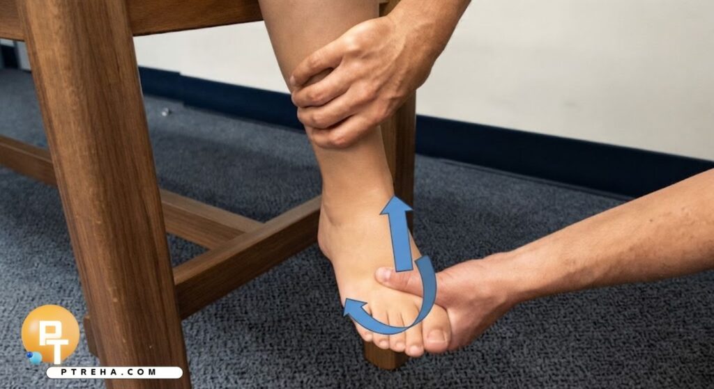
The Kleiger Test is an orthopedic special test used during clinical examination of the ankle, most commonly as an external-rotation stress maneuver when syndesmotic injury is suspected.
The test may reproduce pain associated with injured syndesmotic structures, while some descriptions also discuss medial ankle symptoms related to the deltoid ligament.
Because protocols and terminology vary, the Kleiger Test should be interpreted as one component of a broader ankle assessment rather than as a stand-alone diagnostic test.
What Is the Kleiger Test?
The Kleiger Test is best classified as an orthopedic special test and clinical assessment maneuver for the ankle.
It is commonly described as a manual external-rotation stress test.
The examiner stabilizes the distal lower leg while applying external rotation through the foot, observing whether the maneuver reproduces clinically relevant ankle pain.
Importantly, the terminology is not completely standardized.
A detailed review of the original descriptions and scientific validity of ankle special tests found variation in how sources describe the Kleiger Test, including differences in hand placement, ankle position, and whether the test is discussed primarily in relation to the syndesmosis or deltoid ligament.
For that reason, clinicians should document the actual maneuver performed and the location of symptoms, rather than recording only “Kleiger positive.”
“The location of reproduced pain is as important as the name of the test.”
The Kleiger Test does not independently establish the presence, severity, or stability of a ligament injury.
Purpose of the Kleiger Test
The Kleiger Test may be used as part of an ankle examination to:
- Assess symptoms when a distal tibiofibular syndesmotic injury is suspected.
- Reproduce pain associated with external-rotation loading of the ankle.
- Examine an ankle following a mechanism involving external rotation.
- Help differentiate possible syndesmotic involvement from a more typical lateral ankle sprain.
- Assess whether pain is reproduced around the distal tibiofibular region.
- Observe whether external rotation produces medial ankle symptoms.
- Add information to palpation, injury history, functional examination, and other ankle special tests.
- Help determine whether additional medical or imaging assessment may be appropriate.
It should not be used as the sole method for deciding whether a syndesmotic ligament is torn or whether the ankle is mechanically unstable.
Kleiger Test Indications
The test may be clinically appropriate when the history and examination raise suspicion of a rotational ankle injury.
Possible indications include:
- An ankle injury involving external rotation of the foot relative to the lower leg.
- Pain located over the distal tibiofibular syndesmosis.
- Pain extending proximally from the ankle along the interosseous region.
- Persistent symptoms after an ankle sprain that do not fit a straightforward lateral ligament pattern.
- Difficulty with weight-bearing or push-off associated with suspected syndesmotic injury.
- Medial ankle symptoms following a rotational injury.
- Examination of an athlete with a suspected “high ankle sprain.”
- A need to combine several findings when assessing possible syndesmotic involvement.
The mechanism of injury matters.
Syndesmotic injury can occur with external-rotation forces, but the history alone cannot establish which ligamentous structures are damaged.
Kleiger Test Contraindications and Precautions
There is no universally established list of Kleiger-specific contraindications supported across the clinical literature.
General precautions for provocative ankle testing therefore apply.
The examiner should use particular caution when:
- A fracture is suspected.
- An obvious deformity suggests fracture or dislocation.
- The patient cannot tolerate gentle handling of the ankle.
- Severe acute swelling makes examination unreliable or unnecessarily painful.
- Neurovascular compromise is suspected.
- The injury has not yet been appropriately evaluated following significant trauma.
- External rotation immediately produces substantial pain.
When fracture or serious traumatic injury is possible, appropriate medical evaluation and imaging decisions take priority over aggressive ligament stress testing.
The clinician should not apply progressively greater rotational force simply to obtain a clearer response.
Patient Position
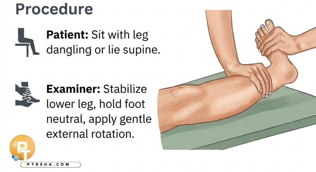
A commonly described manual version of the Kleiger Test uses the following position:
- The patient sits on the examination table.
- The knee is flexed.
- The lower leg is supported or allowed to hang comfortably.
- The ankle is positioned approximately neutral rather than forced into plantarflexion.
- The patient is asked to relax the foot and ankle as much as possible.
Many clinical descriptions use knee flexion of approximately 90 degrees.
A dorsiflexion-external rotation variation is also described in the literature.
This should be documented as a modified protocol rather than assumed to be identical to every version of the Kleiger Test.
Examiner Position
The examiner commonly:
- Positions themselves in front of or slightly lateral to the tested ankle.
- Stabilizes the distal tibia and fibula with one hand.
- Supports or grasps the foot with the other hand.
- Maintains control of the lower leg while rotating the foot relative to it.
- Observes the patient’s response throughout the maneuver.
Exact hand placement varies between published and educational descriptions.
This variation is clinically important because differences in foot position, stabilization, dorsiflexion, and applied force can change the stresses transmitted through the ankle.
How to Perform the Kleiger Test
One commonly used clinical protocol is:
- Position the patient sitting with the knee flexed and the lower leg relaxed.
- Place the ankle in approximately neutral.
- Stabilize the distal lower leg so that the tibia and fibula do not rotate freely with the foot.
- Grasp the foot securely with the opposite hand.
- Explain that the foot will be rotated gently and ask the patient to report the exact location of any symptoms.
- Slowly apply external rotation to the foot relative to the stabilized lower leg.
- Stop if the maneuver produces substantial pain or the patient cannot tolerate further movement.
- Ask the patient where the symptoms occurred rather than recording pain simply as present or absent.
- Relate the finding to palpation, injury mechanism, swelling, functional findings, and other appropriate ankle tests.
The maneuver should be controlled rather than forceful.
A dorsiflexion-external rotation test may place the ankle into dorsiflexion before or during external rotation.
Because this is a protocol modification, findings from different versions should not automatically be treated as interchangeable.
What Constitutes a Positive Kleiger Test?
The terms positive and negative are commonly used for this orthopedic special test, although a positive finding must be interpreted cautiously.
When the test is being used to assess suspected syndesmotic involvement, reproduction of the patient’s characteristic pain around the distal tibiofibular syndesmosis during external rotation is generally considered a clinically relevant positive finding.
Pain may be reported around:
- The anterior distal tibiofibular region.
- The syndesmotic region above the ankle joint.
- The interosseous region between the tibia and fibula.
Some descriptions of the Kleiger Test also consider medial ankle pain relevant when examining potential deltoid ligament involvement.
This difference is one reason documentation should state both the protocol and symptom location.
A positive Kleiger Test does not prove that a particular ligament is completely torn and does not determine whether the syndesmosis is mechanically stable or unstable.
What Does a Positive Kleiger Test Mean?
A positive finding increases clinical suspicion that external-rotation loading is provoking injured or irritated structures around the ankle.
When pain is reproduced over the syndesmotic region, the result may support suspicion of a distal tibiofibular syndesmotic injury when it agrees with the patient’s history and other findings.
It does not establish:
- The exact ligament injured.
- Whether a ligament is partially or completely torn.
- The grade of a syndesmotic injury.
- Whether mechanical instability is present.
- Whether surgical treatment is required.
- Whether another ankle structure is also injured.
A systematic review and meta-analysis of clinical tests for ligamentous injury of the ankle syndesmosis concluded that no individual clinical test demonstrated both high sensitivity and high specificity.
The authors specifically emphasized that clinical tests alone cannot reliably determine injury grade or dictate surgical management.
“A positive stress test contributes evidence; it does not replace the rest of the ankle examination.”
Negative Kleiger Test
A negative Kleiger Test generally means that the maneuver did not reproduce the clinically relevant symptoms expected from the structure or region being assessed.
However, a negative finding should not be interpreted as proof that the syndesmosis is normal.
False-negative clinical findings can occur, and diagnostic performance depends on factors such as:
- Injury severity.
- Acute versus chronic presentation.
- Patient guarding.
- Exact test protocol.
- Examiner technique.
- Location of the injured tissue.
- Presence of other ankle injuries.
If the injury mechanism and other clinical findings continue to suggest syndesmotic injury, further assessment may still be appropriate despite a negative Kleiger Test.
Anatomy Involved in the Kleiger Test
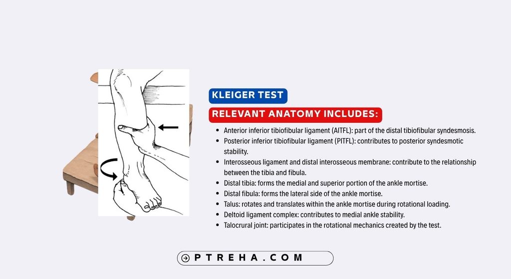
External rotation of the foot relative to the stabilized lower leg can load several structures around the ankle rather than isolating one ligament.
Relevant anatomy includes:
- Anterior inferior tibiofibular ligament (AITFL): part of the distal tibiofibular syndesmosis.
- Posterior inferior tibiofibular ligament (PITFL): contributes to posterior syndesmotic stability.
- Interosseous ligament and distal interosseous membrane: contribute to the relationship between the tibia and fibula.
- Distal tibia: forms the medial and superior portion of the ankle mortise.
- Distal fibula: forms the lateral side of the ankle mortise.
- Talus: rotates and translates within the ankle mortise during rotational loading.
- Deltoid ligament complex: contributes to medial ankle stability.
- Talocrural joint: participates in the rotational mechanics created by the test.
The Kleiger Test should therefore not be described as an isolated test of a single ligament.
External rotation produces a mechanical challenge across several interconnected structures, and the patient’s symptom location must be considered within that broader anatomical context.
Conditions Associated With the Kleiger Test
Findings from the Kleiger Test may be associated with clinical suspicion of:
- Distal tibiofibular syndesmotic sprain.
- Syndesmotic ligament injury.
- So-called high ankle sprain.
- Rotational ankle trauma.
- Deltoid ligament injury when medial ankle symptoms are reproduced.
- Combined ankle ligament injury.
The wording associated with is important.
The test does not independently confirm any of these conditions.
Medial pain during external rotation may raise suspicion involving the medial ankle structures, but physical examination alone cannot reliably establish the extent of deltoid ligament disruption.
A recent systematic review of diagnostic approaches for deltoid ligament injuries demonstrated substantial variation in how deltoid injuries are assessed across studies, including clinical findings, stress radiographs, MRI, ultrasound, and arthroscopy.
Clinical Significance

The main value of the Kleiger Test is as one finding within a clinical assessment, not as an isolated diagnostic decision.
Clinically, the test can:
- Support suspicion of syndesmotic involvement when characteristic pain is reproduced.
- Help relate the patient’s symptoms to an external-rotation mechanism.
- Add information to syndesmotic palpation.
- Complement other ankle special tests.
- Help determine whether the presentation warrants further investigation.
- Encourage examination beyond the lateral ankle ligaments after a rotational ankle injury.
- Help document symptom location and irritability.
- Contribute to decisions about referral or additional diagnostic assessment.
A stronger clinical assessment combines the Kleiger Test with the mechanism of injury, palpation, swelling, weight-bearing tolerance, functional symptoms, other relevant tests, and imaging when indicated.
Sensitivity and Specificity
Diagnostic accuracy for the external rotation test has been studied, but results vary according to population, protocol, reference standard, and study design.
A meta-analysis examining clinical tests for ankle syndesmotic ligament injury pooled four studies of the external-rotation test and reported a specificity of approximately 78% in the included populations, while emphasizing that no individual clinical test demonstrated both high sensitivity and high specificity.
That estimate should not be treated as a universal performance value for every maneuver labeled “Kleiger Test.”
Reasons include:
- Different external-rotation protocols.
- Differences in patient populations.
- Variation in injury severity.
- Different reference standards.
- Variation in ankle positioning.
- Differences between acute and chronic injury assessment.
Older individual studies have also produced substantially different estimates, reinforcing the need to avoid presenting a single number as a universal property of the test.
Diagnostic Accuracy and Limitations
The Kleiger Test has meaningful limitations.
The test cannot reliably determine by itself:
- Whether the syndesmosis is anatomically intact.
- Whether an injury is stable or unstable.
- The exact number of injured ligaments.
- The severity or grade of injury.
- Whether the deltoid ligament is completely disrupted.
- Whether surgical treatment is required.
Another limitation is terminology.
“Kleiger Test,” “external rotation test,” “external rotation stress test,” and “dorsiflexion-external rotation test” are sometimes used interchangeably despite differences in technique.
This creates problems when applying research results to clinical practice.
Evidence obtained with one protocol should not automatically be assumed to describe another.
The clinician should therefore document what was actually done, for example:
External rotation stress test performed with the patient sitting, knee flexed, ankle neutral; external rotation reproduced anterior syndesmotic pain.
That statement communicates considerably more clinical information than simply writing:
Kleiger positive.
Common Mistakes in the Kleiger Test
Common practical errors include:
- Applying external rotation too aggressively.
- Failing to stabilize the distal lower leg.
- Allowing the entire leg to rotate with the foot.
- Recording only “pain” without documenting its location.
- Assuming any ankle pain equals a syndesmotic injury.
- Treating medial pain and syndesmotic pain as equivalent findings.
- Calling the test diagnostic of a ligament rupture.
- Assuming a negative test excludes syndesmotic injury.
- Mixing neutral-position and dorsiflexion protocols without documenting the difference.
- Using a finding from one test to grade the severity of an injury.
- Performing provocative testing before appropriately considering fracture or serious trauma.
- Ignoring findings from palpation, history, function, and other examination procedures.
Another important mistake is interpreting pain intensity as a direct measure of ligament damage.
Greater pain does not automatically mean that a more severe structural lesion is present.
Tips for Performing the Kleiger Test
For a more clinically useful examination:
- Explain the maneuver before performing it.
- Begin with gentle external rotation.
- Stabilize the tibia and fibula consistently.
- Use the same protocol when comparing serial examinations.
- Ask the patient to point to the exact site of reproduced pain.
- Differentiate syndesmotic pain from medial, lateral, posterior, or generalized ankle discomfort.
- Document whether the ankle was neutral or dorsiflexed.
- Document whether symptoms reproduced the patient’s familiar complaint.
- Compare with the opposite side when clinically appropriate, while remembering that side-to-side comparison does not establish pathology by itself.
- Combine the result with syndesmotic palpation and other appropriate clinical findings.
- Stop the maneuver if substantial pain develops.
- Do not increase force simply to produce a positive response.
When teaching the test, it is particularly helpful to emphasize anatomical symptom localization rather than simply demonstrating the foot movement.
Kleiger Test in the Complete Ankle Examination
The Kleiger Test should rarely be interpreted in isolation.
A clinically appropriate ankle assessment may also consider:
- Injury mechanism.
- Location of pain.
- Ability to bear weight.
- Swelling and bruising.
- Palpation of relevant bony and soft-tissue structures.
- Syndesmotic tenderness.
- Ankle range of motion.
- Other syndesmotic clinical tests where appropriate.
- Lateral ligament findings.
- Medial ankle findings.
- Functional tolerance.
- Neurovascular findings when relevant.
- Need for radiographs or other imaging.
The importance of each component depends on the clinical presentation.
For example, a patient with a twisting injury, tenderness over the distal syndesmosis, pain during weight-bearing, and characteristic pain reproduced by external rotation presents a different clinical picture from someone who reports only nonspecific discomfort during the Kleiger Test.
Clinical reasoning comes from the pattern of findings, not from one isolated maneuver.
Summary
- The Kleiger Test is an orthopedic special test involving external rotation of the foot relative to the stabilized lower leg.
- It is commonly used as part of the assessment of suspected distal tibiofibular syndesmotic injury, while some descriptions also relate medial ankle pain to possible deltoid ligament involvement.
- Reproduction of characteristic syndesmotic pain can support clinical suspicion, but the test does not independently diagnose, grade, or determine the stability of a syndesmotic injury.
- Neutral-position and dorsiflexion-external rotation protocols both exist, so clinicians should document the technique they actually use.
- Diagnostic evidence supports interpreting the Kleiger Test together with history, palpation, other examination findings, and appropriate imaging rather than using it as a stand-alone decision tool.
The Kleiger Test can be a useful part of a structured ankle examination when it is performed gently and interpreted anatomically.
Its greatest clinical value comes from identifying whether external-rotation loading reproduces the patient’s characteristic symptoms and then integrating that information with the rest of the examination.