
Kernig Sign is a neurological clinical sign of meningeal irritation used during the examination of a patient in whom meningitis or another process affecting the meninges is suspected.
It is not a stand-alone diagnostic test: the finding must be interpreted alongside the history, neurological examination, systemic findings, and appropriate medical investigations.
What Is the Kernig Sign?
The Kernig Sign is a neurological clinical assessment, more specifically a traditional meningeal irritation sign.
It is not an orthopedic special test, functional performance test, outcome measure, or quantitative measurement.
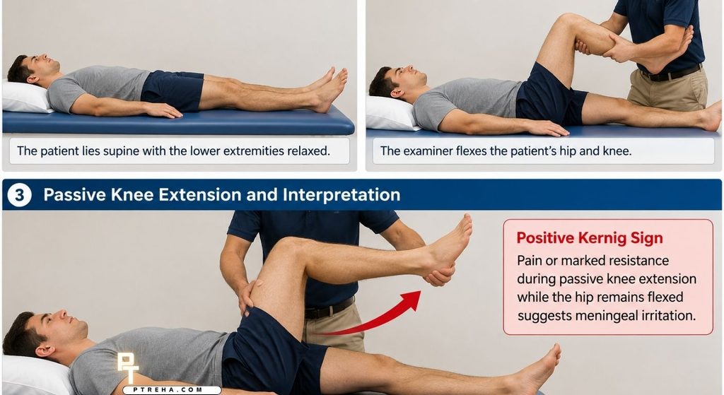
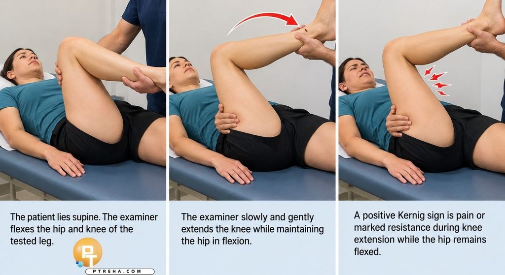
In the commonly described modern technique, the patient lies supine while the examiner flexes the hip and knee and then attempts to extend the knee.
Pain or marked resistance during knee extension constitutes the traditional positive finding.
The proposed basis of the maneuver is that positioning the lower extremity in this way increases tension within neural and meningeal tissues.
When the meninges are inflamed, that maneuver may produce pain or involuntary resistance.
The sign has historically been associated with meningitis, but it should not be used by itself to diagnose or exclude meningitis.
Current WHO guidance on meningitis emphasizes that classical meningeal signs, including Kernig and Brudzinski signs, have limited sensitivity and that their absence cannot rule out meningitis.
“Kernig Sign is a clinical clue to meningeal irritation, not a diagnosis in itself.”
Purpose of the Kernig Sign
Kernig Sign may be incorporated into a neurological examination when meningeal irritation is clinically suspected.
Its purposes include:
- Assessing for a traditional physical sign associated with meningeal irritation.
- Contributing to the examination of a patient with possible meningitis.
- Identifying pain or resistance produced during the characteristic limb maneuver.
- Supporting clinical reasoning when considered with other neurological and systemic findings.
- Complementing assessment for neck stiffness and other meningeal signs.
- Helping determine whether urgent medical evaluation is necessary when the overall clinical presentation is concerning.
- Providing one component of a broader neurological examination rather than establishing a diagnosis independently.
For physical therapists, the main clinical relevance is recognizing that a concerning neurological or systemic presentation requires medical assessment rather than continued musculoskeletal treatment.
Kernig Sign Indications
Kernig Sign may be considered during a medical or neurological examination when the patient’s overall presentation raises concern for meningeal inflammation.
Examples include a patient with combinations of:
- Acute severe headache.
- Fever or a recent febrile illness.
- Neck stiffness.
- Photophobia.
- Nausea or vomiting associated with neurological symptoms.
- Altered mental status.
- Unexplained neurological deterioration.
- Other findings raising suspicion of meningitis or central nervous system infection.
These findings are not interchangeable, and their absence does not necessarily exclude meningitis.
In infants, older adults, immunocompromised people, and some seriously ill patients, the clinical presentation may be atypical.
Kernig Sign therefore has particularly limited value when considered in isolation.
Kernig Sign Contraindications and Precautions
There are no universally established absolute contraindications specific to the Kernig Sign itself.
Clinical judgment should determine whether attempting the maneuver is appropriate.
Relevant precautions include:
- Do not allow the maneuver to delay urgent medical assessment when meningitis is suspected.
- Avoid forceful knee extension.
- Stop if the maneuver produces substantial pain or distress.
- Use caution when significant hip or knee injury limits safe positioning.
- Consider recent surgery, fracture, severe arthritis, or other conditions that make passive hip or knee movement inappropriate.
- Do not repeatedly provoke symptoms merely to reproduce the finding.
- Interpret guarding cautiously because musculoskeletal pain may limit the maneuver.
- Consider whether reduced consciousness, agitation, or inability to cooperate makes examination unreliable.
- Follow appropriate infection-control precautions when an infectious disease is suspected.
These are primarily general examination and safety considerations, not specific diagnostic contraindications to Kernig Sign.
A patient with suspected bacterial meningitis requires urgent medical management.
Performing a bedside meningeal sign should never become a reason to delay escalation of care.
Patient Position
A commonly used modern protocol involves the following patient position:
- Patient lies supine.
- The examined lower extremity is initially relaxed.
- The examiner flexes the hip.
- The knee is also flexed.
- The thigh is maintained in the flexed position while knee extension is attempted.
Descriptions of the exact amount of hip and knee flexion vary among educational and research protocols.
Therefore, the technique should be documented rather than assuming every examiner uses precisely the same method.
Historically described versions of Kernig’s observation were not identical to the contemporary supine maneuver, which is another reason to document the protocol being used.
Examiner Position
The examiner generally stands beside the limb being assessed.
The examiner should:
- Support the patient’s thigh and lower leg.
- Control both hip and knee positioning.
- Keep the patient’s movements passive unless otherwise required.
- Observe for pain, guarding, resistance, and the patient’s overall response.
- Avoid rapid or forceful extension.
- Compare the patient’s response with the broader clinical presentation rather than treating the maneuver as an isolated pass/fail test.
Appropriate support of the leg improves patient comfort and reduces unnecessary muscular effort.
How to Perform the Kernig Sign
A commonly described contemporary procedure is:
- Position the patient comfortably in supine.
- Explain that you will move the patient’s leg passively.
- Flex the patient’s hip and knee.
- Support the thigh so that the hip remains flexed.
- Slowly begin extending the knee.
- Observe for pain, pronounced resistance, or an inability to comfortably continue knee extension.
- Stop if significant discomfort occurs.
- Document the response and the technique used.
- Interpret the finding together with the patient’s neurological and systemic examination.
The examiner should not force the knee through resistance.
The maneuver should also not be reduced to a numerical range-of-motion test.
Although historical descriptions included particular knee angles, a universal modern diagnostic cutoff should not be assumed across patients and protocols.
What Constitutes a Positive Kernig Sign?
The terminology positive Kernig Sign is clinically established and therefore appropriate.
A positive finding is generally described as pain or marked resistance during passive knee extension while the hip is maintained in flexion.
Some descriptions emphasize pain, whereas others emphasize resistance or restriction of extension.
These differences reinforce the importance of documenting what was actually observed.
A finding should not automatically be labeled positive simply because:
- The patient has tight hamstrings.
- Full knee extension is unavailable for orthopedic reasons.
- The patient voluntarily contracts the muscles.
- Hip or knee arthritis causes pain.
- A previous injury restricts motion.
- Neural mechanosensitivity from another cause reproduces symptoms.
The clinical context is essential.
What Does a Positive Kernig Sign Mean?
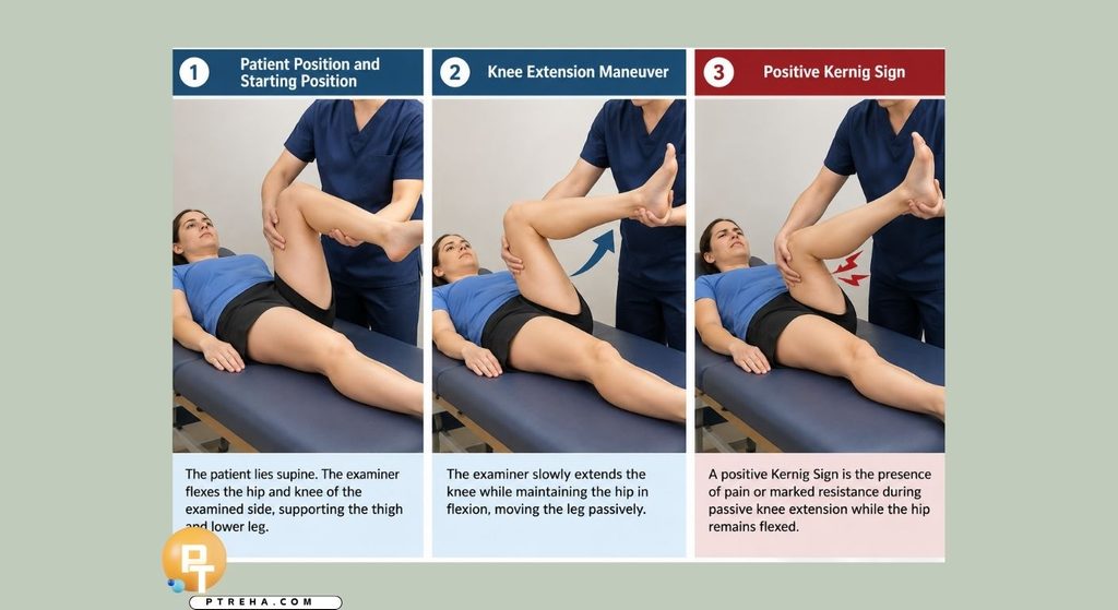
A positive Kernig Sign is traditionally interpreted as evidence that may be consistent with meningeal irritation.
Meningeal irritation can occur with meningitis, but a positive Kernig Sign does not establish:
- The presence of bacterial meningitis.
- The infectious organism.
- The severity of meningitis.
- The absence or presence of intracranial complications.
- The need for a specific treatment by itself.
A positive result should increase clinical attention when it appears in an appropriate clinical picture, especially when accompanied by symptoms such as fever, headache, neck stiffness, altered consciousness, or other neurological abnormalities.
The definitive evaluation of suspected meningitis depends on medical assessment and appropriate investigations rather than the Kernig Sign alone.
Negative Kernig Sign
A negative Kernig Sign means that the characteristic pain or resistance is not reproduced with the maneuver being used.
However, a negative Kernig Sign cannot safely exclude meningitis.
This is one of the most important limitations of the examination.
Many patients with meningitis do not demonstrate classical meningeal signs.
Accordingly:
- Do not dismiss meningitis solely because Kernig Sign is absent.
- Do not use the test as a screening tool capable of ruling out meningitis.
- Continue clinical assessment when symptoms remain concerning.
- Escalate care based on the complete clinical picture.
“The absence of Kernig Sign should never overrule a clinical presentation that remains concerning for meningitis.”
Anatomy Involved in Kernig Sign
Kernig Sign does not selectively test a single anatomical structure.
Relevant anatomy and physiological systems include:
- Meninges: The dura mater, arachnoid mater, and pia mater surround the brain and spinal cord.
- Spinal nerve roots: Movement of the lower extremity can influence neural tissue mechanics.
- Lumbosacral neural structures: Hip flexion combined with knee extension changes the mechanical environment of the neural system.
- Spinal canal: Neural and meningeal tissues travel within the vertebral canal.
- Hip joint: Hip positioning forms part of the examination maneuver.
- Knee joint: Passive knee extension provides the principal provocative movement.
- Hamstring muscles: Muscle length and guarding can influence the apparent resistance encountered.
- Central nervous system: Meningitis involves inflammation of tissues surrounding the brain and spinal cord.
The examination should therefore not be described as a test that “isolates” the meninges.
Musculoskeletal and neural factors can affect the patient’s response.
Conditions Associated With the Kernig Sign
Kernig Sign is most strongly associated clinically with meningeal irritation.
It may be encountered in the assessment of:
- Suspected meningitis.
- Bacterial meningitis.
- Viral or aseptic meningitis.
- Tuberculous meningitis.
- Other conditions that produce meningeal inflammation or irritation.
The finding should be described as associated with these conditions rather than as proof that any particular disorder is present.
The evidence review informing contemporary meningitis guidance also found that the diagnostic performance of Kernig Sign varies according to population and study design.
In adults, combinations involving Kernig or Brudzinski signs may be relatively specific but insufficiently sensitive to exclude disease when absent.
Clinical Significance
The greatest clinical value of Kernig Sign is as one piece of information within a larger neurological and medical assessment.
Clinically:
- A positive finding may increase suspicion of meningeal irritation.
- The result should be interpreted alongside symptoms and other examination findings.
- A negative finding does not reliably rule out meningitis.
- The test should not replace appropriate diagnostic investigations.
- Results may be affected by pain, mobility restriction, guarding, and patient cooperation.
- Classical meningeal signs may be absent even in clinically important meningitis.
- An atypical presentation does not eliminate the possibility of serious disease.
- Suspected central nervous system infection requires prompt medical evaluation.
For physical therapists, recognition is particularly important when a patient initially presents with what appears to be neck pain, headache, spinal pain, or generalized musculoskeletal symptoms.
Fever, significant headache, altered mental state, neurological deterioration, unexplained neck stiffness, systemic illness, or other concerning findings should prompt consideration of a non-musculoskeletal cause and appropriate referral.
Sensitivity and Specificity

The diagnostic performance of Kernig Sign is not sufficiently consistent to justify treating it as an independent diagnostic or exclusion test.
A systematic review and meta-analysis of nine studies evaluating physical meningeal signs reported pooled/provisional estimates for Kernig Sign of approximately 22.9% sensitivity and 91.2% specificity across the included study populations.
The authors emphasized substantial differences among studies and found that Kernig and Brudzinski signs tended to have lower sensitivity and higher specificity than nuchal rigidity or jolt accentuation.
These figures come from the populations and reference standards represented in the included studies and should not be treated as universal values for every patient or clinical setting.
The full systematic review of meningeal signs concluded that a substantial proportion of patients with meningitis do not demonstrate typical meningeal findings on physical examination.
Practically, this means the absence of Kernig Sign has limited value for excluding meningitis.
Diagnostic Accuracy
Kernig Sign has limited stand-alone diagnostic accuracy because its relatively low sensitivity means that many patients with meningitis may not demonstrate the sign.
A positive finding can contribute useful evidence in the correct clinical setting, particularly because specificity has tended to be higher than sensitivity in published studies.
However, the magnitude of that value depends on:
- The population being examined.
- The underlying prevalence of meningitis.
- The reference standard used by the study.
- How Kernig Sign was performed and defined.
- Patient age.
- Examiner technique.
- The underlying cause of meningeal irritation.
- The stage of the illness.
It is therefore inappropriate to interpret Kernig Sign as a definitive diagnostic test.
The diagnosis of meningitis depends on the broader clinical picture and appropriate medical investigations, which may include cerebrospinal fluid assessment and other laboratory or imaging procedures when clinically indicated.
Common Mistakes in Kernig Sign
Several errors can reduce the usefulness of the examination.
Treating Hamstring Tightness as a Positive Sign
Limited knee extension alone does not necessarily indicate meningeal irritation.
Consider:
- Baseline flexibility.
- Chronic knee flexion contracture.
- Previous injury.
- Arthritis.
- Pain from the hip or knee.
- Voluntary guarding.
Extending the Knee Too Quickly
Rapid movement may provoke pain or muscular guarding unrelated to meningeal irritation.
Use slow, controlled passive movement.
Forcing Through Resistance
Kernig Sign does not require the examiner to force maximal knee extension.
Stop when clinically meaningful pain or marked resistance is encountered.
Using the Test to Rule Out Meningitis
This is one of the most clinically important errors.
An absent Kernig Sign does not provide adequate reassurance when the history and examination remain concerning.
Ignoring the Patient’s Overall Presentation
The test should never be interpreted independently from:
- Vital signs.
- Headache history.
- Mental status.
- Neck symptoms.
- Neurological examination.
- Skin findings.
- Recent illness.
- Immune status.
- Overall clinical appearance.
Assuming Every Protocol Is Identical
Historical and contemporary descriptions of Kernig Sign differ.
Document how the test was performed rather than assuming that the label alone fully describes the maneuver.
Tips for Performing the Kernig Sign
For a clinically meaningful examination:
- Explain the maneuver before beginning.
- Position the patient comfortably.
- Keep the patient as relaxed as possible.
- Support the thigh and lower leg.
- Move slowly.
- Maintain control of hip position.
- Observe rather than simply asking whether the movement hurts.
- Note whether resistance appears voluntary or involuntary.
- Consider musculoskeletal causes of restricted knee extension.
- Avoid assigning significance to a small difference in range of motion.
- Document the patient’s specific response.
- Record relevant neurological and systemic findings.
- Never use a negative Kernig Sign to dismiss an otherwise concerning presentation.
- Prioritize urgent medical referral when meningitis remains clinically possible.
Kernig Sign in Physical Therapy Practice
Kernig Sign is not typically used to direct musculoskeletal rehabilitation or prescribe an exercise program.
Its relevance to physical therapy is primarily recognition and referral.
For example, suppose a patient presents for treatment of new neck pain but also reports severe headache and systemic illness.
If neurological assessment raises concern for meningeal irritation, continuing routine cervical mobility exercises would not be the appropriate priority.
The therapist should instead recognize that the presentation may fall outside ordinary musculoskeletal management and arrange appropriate medical evaluation according to the urgency of the presentation.
Similarly, a patient who does not demonstrate Kernig Sign may still have serious neurological or infectious pathology.
Screening decisions should therefore never depend on this maneuver alone.
Summary
- Kernig Sign is a neurological clinical sign used to assess for findings associated with meningeal irritation.
- The commonly used technique involves hip and knee flexion followed by controlled passive knee extension.
- Pain or pronounced resistance during knee extension constitutes the traditional positive finding.
- A positive Kernig Sign may support suspicion of meningeal irritation but does not independently diagnose meningitis.
- A negative Kernig Sign does not reliably exclude meningitis, making the complete clinical presentation and appropriate medical investigation essential.
Kernig Sign remains useful as part of neurological examination and clinical reasoning, but its limitations are as important as the maneuver itself.
In physical therapy practice, the key responsibility is recognizing a potentially serious non-musculoskeletal presentation and ensuring that bedside testing does not delay appropriate medical assessment.