Homans Sign is a historical physical examination maneuver associated with assessment of suspected lower-extremity deep vein thrombosis (DVT).
It describes calf discomfort produced during passive ankle dorsiflexion, but the finding is nonspecific and should not be used to diagnose or rule out DVT.
In current physical therapy practice, its main value is educational: clinicians should recognize the sign while understanding that suspected DVT requires appropriate clinical risk assessment and medical diagnostic testing.
What Is the Homans Sign?
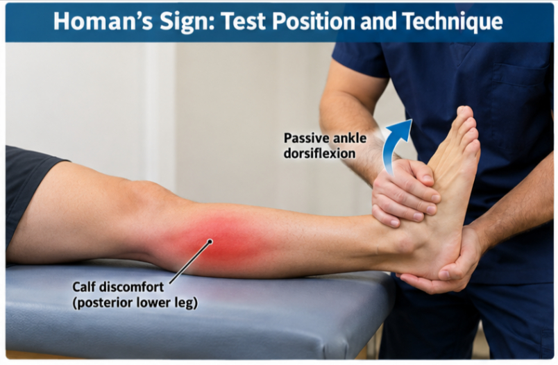
Homans Sign, also written as Homan’s sign, is a clinical physical examination sign historically used when evaluating a patient for possible lower-extremity DVT.
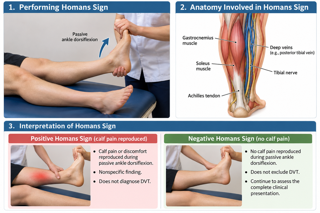
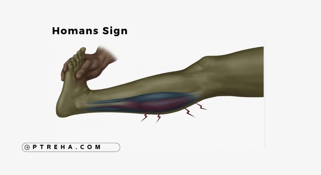
In the commonly described maneuver, the examiner passively dorsiflexes the patient’s ankle.
Reproduction of calf pain has traditionally been called a positive Homans Sign.
However, this response does not demonstrate the presence of a venous thrombus.
Calf pain during ankle dorsiflexion can arise from multiple musculoskeletal and other conditions.
From a modern clinical perspective, Homans Sign is therefore best classified as a historical clinical assessment sign rather than a reliable diagnostic or stand-alone screening test.
The distinction matters.
A physical therapist should never reassure a patient that DVT is absent simply because Homans Sign does not reproduce pain.
The broader evidence supports this caution.
The American Society of Hematology diagnostic guideline for venous thromboembolism recommends diagnostic strategies based on pretest probability and appropriate laboratory or imaging testing rather than relying on an isolated physical sign.
“Homans Sign is a clinical finding, not a diagnosis.”
Purpose of the Homans Sign
Historically, clinicians used Homans Sign as part of the physical examination when DVT was suspected.
Its potential purposes have included:
- Observing whether passive ankle dorsiflexion reproduces calf pain.
- Contributing historically to examination of an acutely painful or swollen calf.
- Recognizing a traditional physical examination finding associated with suspected DVT.
- Teaching clinicians why isolated signs have limited diagnostic value.
- Encouraging consideration of vascular causes when calf symptoms cannot be explained adequately by the musculoskeletal examination.
- Supporting broader clinical reasoning when viewed alongside history, symptoms, risk factors, and other findings.
It should not be used for:
- Confirming DVT.
- Excluding DVT.
- Determining whether medical referral is unnecessary.
- Deciding that exercise or manual therapy is safe when significant DVT suspicion remains.
- Replacing an established clinical prediction approach.
- Replacing D-dimer testing or diagnostic imaging when these are clinically indicated.
In practice, the most important lesson is not how strongly the maneuver reproduces symptoms but whether the patient’s overall presentation suggests that a potentially serious vascular condition requires medical assessment.
Homans Sign Indications
There is no strong contemporary indication to perform Homans Sign routinely as a diagnostic maneuver for DVT.
A clinician may encounter or discuss it when:
- Reviewing a historical vascular examination.
- Teaching physical therapy students about DVT assessment.
- Interpreting documentation from another clinician.
- Examining a patient whose previous medical record states that Homans Sign was positive or negative.
- Differentiating older examination practices from current evidence-based pathways.
- Considering vascular pathology within the differential assessment of unexplained calf pain or swelling.
When a patient presents with findings that raise meaningful concern for DVT, the priority is appropriate risk assessment and referral rather than repeatedly provoking calf symptoms to obtain Homans Sign.
Clinical suspicion may arise from the overall pattern rather than one finding—for example, otherwise unexplained unilateral swelling, pain, tenderness, skin changes, relevant recent immobilization or surgery, previous venous thromboembolism, active malignancy, or other established risk factors.
The precise significance of these features depends on the clinical context and the validated prediction strategy being used.
Homans Sign Contraindications and Precautions
There is not a universally established list of test-specific contraindications for Homans Sign comparable with those used for some orthopedic special tests.
More importantly, the maneuver has limited diagnostic usefulness, so there is generally little reason to provoke symptoms merely to obtain the sign when DVT is already a realistic clinical concern.
Practical precautions
- Do not use Homans Sign to determine whether a patient is safe to continue rehabilitation.
- Do not repeatedly force ankle dorsiflexion in an acutely painful or swollen calf.
- Avoid aggressive movement when the patient has significant acute pain.
- Do not allow a negative result to override concerning symptoms or risk factors.
- Do not postpone medical assessment while attempting multiple physical tests for DVT.
- Follow local referral and emergency procedures when clinically significant DVT or pulmonary embolism is suspected.
- Consider recent trauma, surgery, immobilization, neurological impairment, and musculoskeletal injuries when deciding whether passive ankle movement is appropriate.
- Stop the examination if the patient’s condition suggests an urgent medical problem outside the scope of musculoskeletal treatment.
For physical therapists, the key safety issue is clinical triage, not obtaining the Homans Sign.
A patient with concerning findings should be managed according to the appropriate vascular assessment and referral pathway.
Patient Position
Descriptions of Homans Sign are not completely uniform across historical sources.
A commonly described position includes:
- Patient lying supine.
- Lower extremity supported and relaxed.
- Knee commonly maintained relatively extended in traditional descriptions.
- Ankle initially in a comfortable position.
- Patient instructed to report calf discomfort during the movement.
Because different descriptions exist, the exact position used should be documented rather than presented as a universally standardized protocol.
The patient’s resting symptoms should also be noted before movement.
Calf pain that is already severe at rest should not be interpreted as being specifically produced by the maneuver.
Examiner Position
The examiner typically positions themselves so the ankle can be moved gently without unnecessary force.
The examiner may:
- Stand near the patient’s foot.
- Support the lower extremity as required.
- Stabilize the leg without forcefully compressing the calf.
- Hold the foot or ankle securely.
- Move the ankle into dorsiflexion gradually.
- Observe the patient’s response.
- Ask where any reproduced discomfort is located.
The maneuver is not intended to be an aggressive calf stretch.
Forceful dorsiflexion provides no reliable additional diagnostic information and may simply increase musculoskeletal discomfort.
How to Perform the Homans Sign
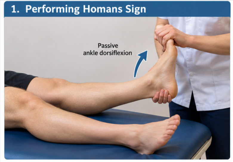
A traditional description of the maneuver is:
- Position the patient comfortably, usually in supine.
- Allow the involved leg to relax.
- Establish whether calf pain is already present at rest.
- Support the leg appropriately.
- Hold the foot or ankle.
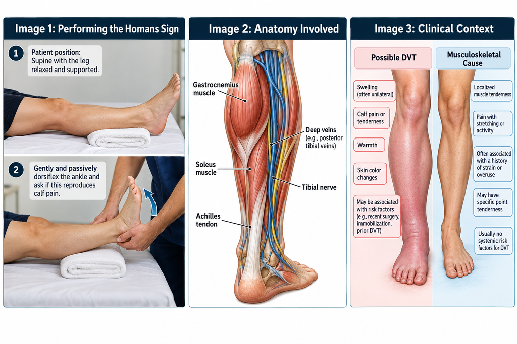
- Gently passively dorsiflex the ankle.
- Ask whether the maneuver produces or increases calf discomfort.
- Note the location and character of any symptoms.
- Stop if movement causes substantial discomfort.
- Interpret the response only in the context of the entire clinical presentation.
The traditional maneuver may appear straightforward, but its simplicity should not be confused with diagnostic accuracy.
An older study examining clinical symptoms and signs found that classic findings—including Homans Sign—occurred with similar frequency in limbs with and without venographically demonstrated DVT, illustrating why the sign cannot reliably distinguish the two groups.
For a physical therapist, performing several repetitions to see whether the symptom becomes clearer is not clinically useful.
What Constitutes a Positive Homans Sign?
The term positive Homans Sign is historically appropriate because this examination maneuver has traditionally been documented using positive and negative terminology.
A positive finding generally refers to:
- Calf pain or discomfort reproduced during passive ankle dorsiflexion.
However, this definition describes only the response to the maneuver.
It does not mean:
- DVT has been diagnosed.
- A blood clot has been located.
- A specific vein has been identified as thrombosed.
- The calf pain originates from a vein.
- Anticoagulation is indicated.
- Imaging is automatically positive.
Pain with ankle dorsiflexion may occur because the maneuver also moves or tensions musculoskeletal tissues in and around the calf.
For this reason, the finding is inherently nonspecific.
What Does a Positive Homans Sign Mean?
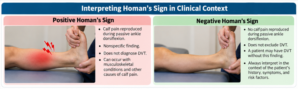
A positive Homans Sign means that passive ankle dorsiflexion reproduced calf discomfort during the particular examination.
That is the most that can safely be concluded from the sign itself.
A positive response may be seen in a patient who has DVT, but it can also occur in patients without DVT.
Potential alternative explanations for calf pain during dorsiflexion can include musculoskeletal disorders involving the calf or ankle and other conditions capable of producing a painful or swollen lower leg.
The examiner therefore should ask a more clinically useful question:
Does the complete history and examination create sufficient suspicion of DVT to require an appropriate medical diagnostic pathway?
Modern DVT evaluation uses the patient’s clinical probability together with appropriate testing.
A systematic review supporting contemporary diagnostic strategies found substantially better evidence for established investigations such as compression ultrasonography and D-dimer testing than for isolated clinical signs.
The systematic review of diagnostic testing for lower-extremity DVT explains how these tests function within diagnostic pathways rather than as isolated examination signs.
“When DVT is a realistic concern, the next decision is whether the patient needs medical investigation—not whether another calf maneuver can reproduce pain.”
Negative Homans Sign
A negative Homans Sign generally means that passive ankle dorsiflexion did not reproduce the traditional calf-pain response.
It does not mean DVT has been excluded.
This is one of the most important clinical teaching points associated with the sign.
A patient may have DVT without showing calf pain during ankle dorsiflexion.
Therefore:
- Do not document “DVT ruled out” because Homans Sign is negative.
- Do not reduce appropriate referral solely because the maneuver is painless.
- Do not resume exercise based only on a negative Homans Sign when other concerning findings remain.
- Continue considering the complete history and clinical presentation.
- Use an appropriate clinical decision pathway when DVT is suspected.
This limitation is especially important in rehabilitation because patients may initially present to a physical therapist for what appears to be musculoskeletal calf pain.
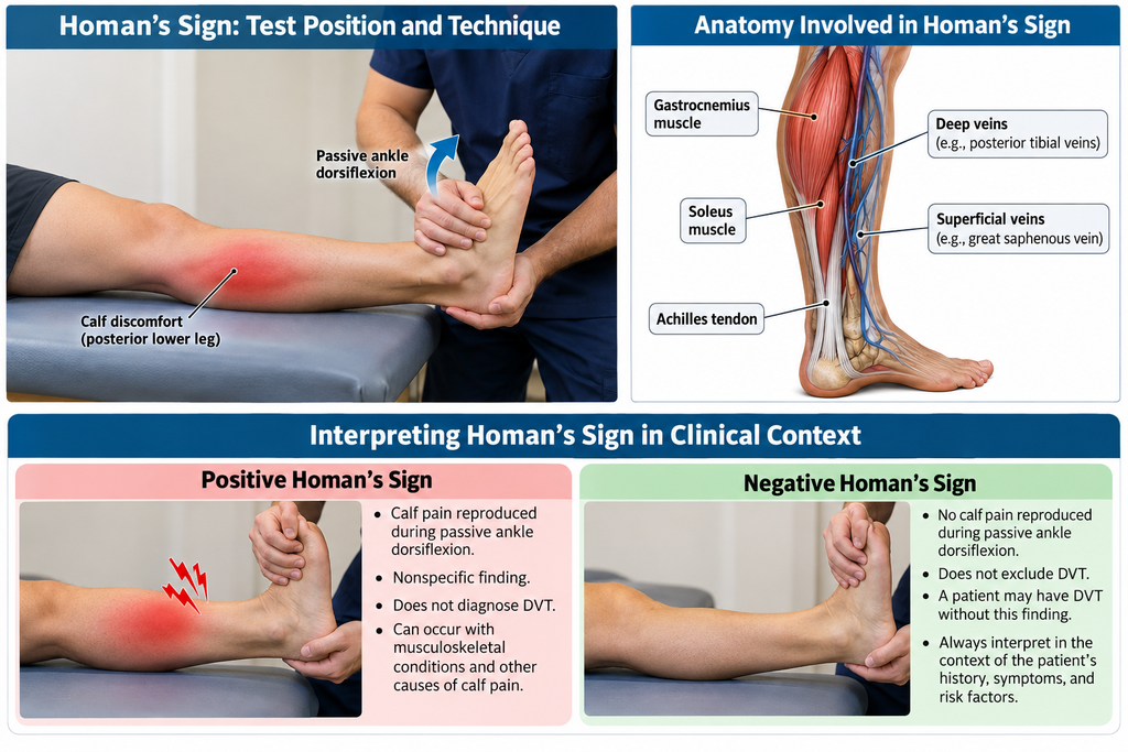
Anatomy Involved in Homans Sign
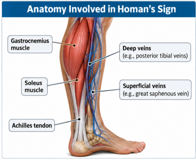
Homans Sign does not isolate one vein, muscle, tendon, or other anatomical structure.
Passive ankle dorsiflexion affects several tissues and mechanical relationships throughout the posterior lower leg.
Relevant anatomy includes:
- Gastrocnemius muscle.
- Soleus muscle.
- Achilles tendon and associated musculotendinous structures.
- Deep posterior compartment tissues.
- Superficial and deep venous systems of the lower extremity.
- Tibial and fibular structures surrounding the calf.
- Ankle joint and periarticular tissues.
- Neural structures traveling through the posterior leg.
The gastrocnemius crosses both the knee and ankle, meaning its mechanical response to ankle dorsiflexion is influenced by knee position.
The soleus crosses the ankle but not the knee.
These relationships help explain an important limitation: pain during passive dorsiflexion is not inherently vascular.
A painful calf muscle, healing strain, irritated musculotendinous tissue, or another local disorder may also become symptomatic during the movement.
Homans Sign therefore cannot identify which anatomical structure is responsible for the patient’s pain.
Conditions Associated With Homans Sign
The condition historically associated with Homans Sign is:
- Lower-extremity deep vein thrombosis.
However, a painful response is nonspecific and may occur in other painful lower-leg presentations.
Clinical differential assessment may therefore need to consider conditions such as:
- Calf muscle injury.
- Other musculotendinous calf disorders.
- Local soft-tissue irritation.
- Lower-extremity trauma.
- Cellulitis when the broader clinical presentation is compatible.
- Ruptured or symptomatic popliteal cyst in an appropriate presentation.
- Venous disorders other than acute DVT.
- Other causes of painful unilateral leg swelling.
This is not a list of conditions diagnosed by Homans Sign.
Rather, it demonstrates why reproduction of calf pain cannot identify DVT on its own.
Clinical Significance
For contemporary physical therapy practice, the major clinical significance of Homans Sign lies in understanding its limitations.
Important points include:
- DVT can have serious consequences.
- Individual clinical signs do not reliably establish the diagnosis.
- Homans Sign should not function as a stand-alone screening tool.
- A positive sign is nonspecific.
- A negative sign cannot safely exclude DVT.
- Clinical reasoning should consider symptoms, risk factors, and the overall presentation.
- Validated pretest-probability approaches are more useful than an isolated provocative maneuver.
- Appropriate patients require medical investigation rather than additional musculoskeletal testing.
- Compression ultrasonography is an important component of contemporary DVT diagnostic pathways.
- D-dimer testing is used selectively according to clinical probability and the diagnostic pathway.
This changes the therapist’s role substantially.
The physical therapist is not trying to diagnose the clot through ankle movement.
The therapist is recognizing whether the presentation could represent a vascular problem requiring medical investigation.
Practical example
Consider a patient referred for calf pain after a period of reduced mobility.
The therapist notices new unilateral swelling and calf discomfort that does not fit the expected musculoskeletal pattern.
Performing Homans Sign and finding no pain should not provide reassurance.
Instead, the therapist should consider the entire presentation and follow an appropriate DVT assessment and referral pathway.
The reverse is also true.
If passive ankle dorsiflexion causes calf pain in a patient with a clearly established calf strain, that finding does not automatically indicate DVT.
Clinical context determines the next step.
Sensitivity and Specificity
Reliable contemporary sensitivity and specificity values that could justify using Homans Sign as a stand-alone diagnostic test are not established sufficiently for clinical use.
Published values vary substantially across studies and patient populations, and older studies were conducted using different methods and reference standards.
Presenting one sensitivity or specificity figure as though it were a universal property of Homans Sign would therefore be misleading.
More importantly, contemporary evidence-based DVT diagnostic pathways do not rely on the maneuver.
The practical conclusion is straightforward:
- A positive Homans Sign does not reliably rule in DVT.
- A negative Homans Sign does not reliably rule out DVT.
- Its diagnostic performance is inadequate for independent clinical decision-making.
This is why the therapist should not convert the maneuver into an informal probability estimate such as “DVT unlikely because Homans is negative.”
Diagnostic Accuracy
Homans Sign has poor diagnostic usefulness when considered by itself.
A broader evidence review of clinical findings concluded that no single clinical finding can accurately diagnose DVT.
Modern diagnostic strategies instead combine clinical pretest probability with appropriately selected objective tests.
Depending on the patient’s probability category and clinical circumstances, these may include:
- D-dimer testing.
- Proximal compression ultrasonography.
- Whole-leg ultrasonography.
- Repeat or serial imaging in selected diagnostic pathways.
The exact pathway depends on the clinical setting and patient population.
Consequently, describing Homans Sign as a “DVT test” without qualification can create the wrong clinical expectation.
It is more accurate to describe it as a historical physical sign associated with suspected DVT that lacks adequate accuracy for independent diagnosis or exclusion.
Common Mistakes in Homans Sign
Using it to rule out DVT
This is the most clinically important mistake.
Absence of calf pain during dorsiflexion cannot establish that a patient is free of DVT.
Treating a positive sign as a diagnosis
Pain during dorsiflexion is nonspecific.
A positive maneuver does not confirm venous thrombosis.
Ignoring the patient’s risk profile
The examination should not be interpreted separately from the history.
Relevant risk factors and associated symptoms often matter more than the maneuver itself.
Performing the maneuver repeatedly
Repeated provocative testing does not solve its poor diagnostic accuracy.
Once serious vascular pathology is a realistic concern, further unnecessary symptom provocation adds little useful information.
Using excessive force
Forceful dorsiflexion may simply increase calf or ankle pain.
The historical maneuver involves passive movement, not aggressive stretching.
Assuming calf pain identifies a vein
Passive dorsiflexion stresses several tissues.
The response cannot localize symptoms specifically to the deep venous system.
Treating it like an orthopedic special test
Homans Sign should not be approached like a high-value musculoskeletal test intended to implicate a particular anatomical structure.
Its historical association with DVT does not make it anatomically specific.
Letting the sign delay referral
A therapist should not perform a series of additional orthopedic tests while a concerning vascular presentation goes unaddressed.
Safety and appropriate escalation take priority.
Tips for Physical Therapists
When teaching or encountering Homans Sign in practice:
- Describe it as a historical clinical sign.
- Explain explicitly that it cannot diagnose DVT.
- Document the patient’s symptoms rather than overemphasizing the test label.
- Assess the complete clinical presentation.
- Look for patterns inconsistent with the expected musculoskeletal disorder.
- Consider established DVT risk factors.
- Use validated clinical decision processes when they fall within your setting and scope of practice.
- Follow local clinical pathways for suspected DVT.
- Communicate clearly during referral.
- Report relevant changes such as new swelling, unexplained pain, or other concerning findings.
- Avoid reassurance based only on a negative Homans Sign.
- Avoid diagnosing DVT based only on a positive Homans Sign.
- Escalate appropriately when the presentation suggests a potentially serious vascular condition.
From a practitioner perspective, the safest approach is to recognize when a patient’s presentation has moved beyond routine musculoskeletal management.
Homans Sign in Modern Physical Therapy Practice
Homans Sign is still encountered in textbooks, older clinical documentation, examinations, and discussions of DVT.
That makes understanding it useful.
Using it as a decisive DVT test, however, is not supported by modern evidence.
When teaching students, it is valuable to demonstrate the maneuver primarily to explain how clinical practice has evolved.
Students should understand the distinction between:
- A historically associated physical sign.
- A validated clinical prediction approach.
- A laboratory investigation.
- Diagnostic imaging.
- A confirmed medical diagnosis.
This distinction supports safer clinical reasoning.
Physical therapists frequently assess patients with lower-leg pain, postoperative symptoms, reduced mobility, sports injuries, and swelling.
Some of these patients can have conditions outside the musculoskeletal system.
Recognizing when the clinical pattern warrants referral is therefore much more important than memorizing Homans Sign as a special test.
When Should a Physical Therapist Refer?
Referral should be based on the overall concern for venous thromboembolism, not the Homans Sign alone.
A therapist should consider appropriate medical escalation when the history and presentation raise significant concern for DVT.
Examples of potentially relevant findings can include:
- New unexplained unilateral leg swelling.
- New calf pain or tenderness without a convincing musculoskeletal explanation.
- A meaningful combination of DVT risk factors and lower-extremity symptoms.
- Unexpected deterioration during rehabilitation.
- Symptoms developing after substantial immobilization or another relevant risk exposure.
- A clinical presentation inconsistent with the expected course of the treated condition.
The appropriate urgency depends on the patient’s presentation and local care pathway.
Symptoms suggesting possible pulmonary embolism, such as new unexplained shortness of breath, chest pain, collapse, or other acute cardiopulmonary deterioration, require urgent medical evaluation rather than continued physical therapy examination.
The therapist’s responsibility is appropriate recognition and escalation—not medical treatment of DVT.
Summary
- Homans Sign is a historical clinical sign in which passive ankle dorsiflexion reproduces calf discomfort.
- A positive Homans Sign is nonspecific and does not diagnose DVT.
- A negative Homans Sign does not exclude DVT.
- The maneuver does not isolate the deep venous system because ankle dorsiflexion also affects multiple musculoskeletal tissues.
- Contemporary DVT assessment relies on clinical probability and appropriate objective investigation rather than Homans Sign alone.
Homans Sign remains useful to know because clinicians will continue to encounter the term.
Its most important educational message, however, is its limitation: when DVT is genuinely suspected, safe clinical reasoning and appropriate referral are considerably more important than the result of a calf-provocation maneuver.